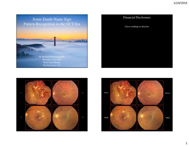

12/4/2015 Financial Disclosures Some Dumb Name Sign Pattern Recognition in the OCT Era I have nothing to disclose H. Richard McDonald MD Brandon Lujan MD West Coast Retina San Francisco, CA 1 2 BVO Drusen 3 4 DME ERM 1
12/4/2015 Sjogren’s Reticular VKH 1 2 Dystrophy Amalric’s Triangular CSR 3 4 Syndrome GCA Pattern Recognition Once you know the pattern, it’s easier to recognize the disease You see what you know Optical Coherence Tomography The more you know, the more you see 2
12/4/2015 Case One Case Two Case Four Case Three 3
12/4/2015 What if you don’t know the pattern? Diagnoses? Courtesy of Christine Curcio, PhD Courtesy of Christine Curcio, Ph.D. 2 Original in IOVS 2011 Case One Nerve Fiber Layer Ganglion Layer Inner Plexiform Layer Inner Nuclear Layer Outer Plexiform Layer Outer Nuclear Layer External Limiting Membrane Ellipsoid Zone (IS/OS) Interdigitation Zone (COST) RPE Normal OCT You have to count the layers 4
12/4/2015 26 year old pregnant woman Severe Fibrinous CSR Transplant patient on prednisone 20/50 The central leak has a geyser-like effect, Batman Sign blowing the fibrin out of the way Fibrinous CSR Fibrin Subretinal fluid 5
12/4/2015 Hemorrhage, perivenular whitening, cotton wool spot associated with non ischemic CRVO Case Two 20/20 En Face OCT through the INL reveals SD-OCT perivenular hyper-reflectivity associated with the ischemia Intraretinal and subretinal fluid 5/200 Patchy INL hyper-reflectivity caused by Deep retinal capillary ischemia 6
12/4/2015 Dash Sign Case Three Paracentral acute middle maculopathy (PAMM): a new variant of acute macular neuroretinopathy associated with retinal capillary ischemia. Sarraf D, et al. JAMA Ophthalmol 2013;131:1275-1287. PAMM is deep capillary ischemia, which is thought to represent a variant of AMN Multimodal imaging findings in retinal deep capillary ischemia. Yu S, et al. Retina 2013:Nov 14. [Epub ahead of print]. PAMM is found in other ischemic etiologies, similar to that seen in our CRVO case Evaluation of cystoid change phenotypes in Ocular Toxoplasmosis using optical coherence tomography Hyper reflectivity caused by Retinitis Ouyang Y, et al., PLoS One 2014;9:e86626 Subretinal fluid with fibrin layer and Eyes with ocular toxoplasmosis may present with HORCS possibly photoreceptor outer segments (Huge Outer Retinal Cystoid Space) 7
12/4/2015 HORCS? Jaws Sign (Huge Outer Retinal Cystoid Space) Really, follow the ELM! The inflammatory process results in exuberant SRF formation causing photoreceptor delamination. The regeneration of PROS may act like a crystal and may be a scaffold for fibrin deposition, resulting in “teeth.” Optical Coherence Tomography Case Four The OCT appears similar in both eyes. HM HM 20/25 20/25 If you don’t know the pattern, you must count the layers! 8
12/4/2015 What’s going on? Our patient has no nerve fiber layer! Normal Normal Our Patient Dull gray Inner Plexiform Layer Bright Inner Plexiform Layer Bright Nerve Fiber Layer NO Nerve Fiber Layer Pituitary Tumor! Tumor resected Vision improved to 20/40 9
12/4/2015 It’s not always what’s there that is the problem Some Dumb Name Sign It may be what’s not there Pattern Recognition in the OCT Era Normal H. Richard McDonald MD Our Patient Brandon Lujan MD West Coast Retina Dog Not Barking Sign! San Francisco, CA 10
Recommend
More recommend