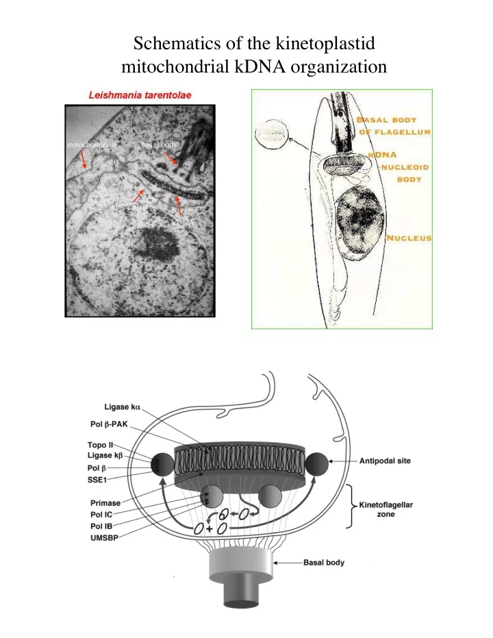

Schematics of the kinetoplastid mitochondrial kDNA organization
And how were these networks labeled In contrast to the ones before, these were labeled in vitro, after the networks were isolated. DNA polymerase or Terminal deoxynucleotidal transferase (TdT) were added to the isolated networks. Both will be active on Nicked or Gapped substrates ( DNA Polymerase can add Labeled nucleotides to the 3’OH and so can TdT. Here, the nucleotide is radio active Here, the nucleotide is fluorescent Same experiment, different label
kDNA rotates during replication …except in T brucei, where it oscillates
How are these kDNA networks labeled? Two methods: In vivo: PULSE/CHASE In vitro: Labeling nicked and gapped Minicircles In this figure, the in vivo labeling trick was used: Cells are grown in the presence of a radioactive Thymidine for A) 3 min B) 6 min C) 30 min and D) 60 min (That’s the Pulse). Any DNA replicated during the pulse will incorporate the radioactive Thymidine. A Pulse/chase experiment is when you grow the cells in the presence of the labeled substrate (pulse) and then grow the cells in the presence of unlabeled substrate for a defined period of time after (chase)
Recommend
More recommend