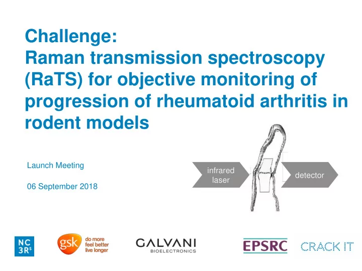

Challenge: Raman transmission spectroscopy (RaTS) for objective monitoring of progression of rheumatoid arthritis in rodent models Launch Meeting infrared detector laser 06 September 2018
Content Introduction to Galvani Bioelectronics • The Challenge • Brief Introduction to Rheumatoid Arthritis (RA) • Current approaches and limitations for RA diagnostics • Overall Goal and proposed approach • Deliverables • GSK/Galvani Panel •
GSK/Galvani Bioelectronics GlaxoSmithKlin Verily Life e Sciences (GSK)
The Challenge A device to evaluate arthritis severity in the rodent joint: • Develop a device capable of detecting the key pathological biomarkers of RA progression • The sensing technology may involve Raman transmission spectroscopy or other spectroscopy techniques • The final device should be hand-held to evaluate the joints in awake rats with minimal animal handling
Key events in the RA pathophysiology synovial inflammation physical X-ray assessment assessment synovial (indirect) (direct) inflammation normal adipose inflammation pannus cartilage/bone damage cartilage damage, joint hypoxia, glycolysis bone damage
Current approaches for RA diagnostics Visual assessment • Published approaches vary according to the degree of (awake animals) animal handling • Tools for RA disease progression are limited, Serology assessment especially for the (awake animals) destruction of bone/cartilage • Push toward non- destructive diagnostics in X-ray, MRI, US, optical awake animals with (anesthetised animals) minimal handling
Progression of arthritis in rat model Pre-ar arthrit thritic ic phase Diseas ase e Diseas ase e 16 Loss of immune tolerance Progres ression ion Onset al Score re 12 X-ray ray ical 8 Clinic 4 0 0 7 15 21 Primary immunisation Booster immunisation Type II collagen/adjuvant Type II collagen/adjuvant (i.d.) Day (i.d.) • Clinically apparent arthritis with swollen joints appears 12 – 14 days after the primary immunisation. • Animals reach maximum severity by day 21 (bone changes)
Current approaches for RA diagnostics • Body weights (PPL severity limit: -20% bodyweight loss) • Visual Assessment – Clinical scores/welfare parameters • Paw Volumes – Plethysmometer measurement • Imaging – MRI / Optical imaging • Possible Serology – Intraveneous blood sampling via tail vein. • Terminal Sampling - Imaging: CT scanner - Histological analysis of hind limbs - Sections combined and processed for gene analysis. - Serology: serum for antibodies, proteins and cytokines.
Visual Assessment - typical rat paw appearance X-ray ray Figure 1. Comparison of rats (A) before and (B) after modelling CIA. Human Figure 2. The joint inflammation which develops in rodent arthritis models (CIA) resembles inflammation in human patients with RA.
Visual Assessment - typical mouse paw appearance Methods for visually scoring or quantifying the amount of joint X-ray ray inflammation; these are semi- quantitative at best and subject to significant inter-observer variability. CIA is scored blind, by a person unaware of both treatment and of previous scores for each animal. Extensive training and practice is critical to repeatable scoring. The animal's score is the total of all four paw scores on scale of 0-16, as shown.
Paw Volumes – Plethysmometer Measurement The Ugo Basile™ Plethysmometer is a microprocessor-controlled volume X-ray ray meter that has long been the standard instrument for measurement of rodent paw volume. The first device designed specifically to measure paw swelling in rodents. More than 1000 bibliographic citations since 1960s. It consists of a water filled Perspex cell into which the rat paw is dipped. A transducer records small differences in water level caused by volume displacement, operates a graphic LCD read-out which shows the exact volume of the paw (control or treated).
Paw Volumes – Plethysmometer Measurement Advantages: • The Plethysmometer enables a rapid X-ray ray screening of a large number of rats; • The inflammation is quantifiable; • Evaluation of small volume differences • Comfortable reading on the graphic display • Data recordings are digital. Direct connection to: PC and Mini Printer. Disadvantages: • Depth in which the paw is introduced can be different; • The moment in which the measurement of inflammation is taken may not be the same, human error.
Imaging techniques for arthritis diagnostics MRI X-ray ray X-ray, MRI, optical imaging, and ultrasound assessment: • slow manual analysis • low diagnostic value for inflammation • no specific chemical information about bone/cartilage damage • X-ray is more readily available but suffers from lower specificity for bone/cartilage damage, compared to MRI, optical imaging & ultrasound
Key drivers for this Challenge: Push toward non-destructive diagnostics in awake animals with minimal handling 3Rs Scientific Patient Challenge Drivers
Patient and Scientific Benefits • RA is characterized by a step-wise progression with multiple relapses, necessitating frequent assessments of the disease severity • Existing diagnostic methods rely on the physical (subjective), X-ray, and serology assessment • No objective specific methods for assessing the bone/cartilage damage Physical assessment X-ray assessment Serology assessment Severity 0 5 10 15 20 25 30 Years of Disease
3Rs Benefits Refinement • shorter study duration and reduced disease severity due to being able to measure the cartilage damage during early RA progression and initial recovery, as compared to the current ability to detect the bone damage (by micro-CT) no earlier than day 21 • as a consequence of the refined study outcomes, more data-rich information is generated regarding the cartilage and bone damage and healing Reduction • smaller number of animals per experimental group due to longitudinal measurements and within-animal repeated-measured design
Overall goal • Develop and validate a handheld device for objective monitoring of RA progression in un-anaesthetised rodents (either restrained or unrestrained). • While it is expected that Raman transmission spectroscopy would be the technology needed to solve the Challenge, other spectroscopy approaches (such as absorption spectroscopy) are also welcome. • Excluded from the challenge: approaches that use transgenic animals or other procedures that substantially complicate the study design (e.g. multiple injections of dyes or nanoparticles).
Proposed approach: in vivo Raman transmission spectroscopy • Selection of optimal infrared laser wavelength for minimal amount of absorption in the skin and bone • Optimisation of the optics for maximal amount of light capture by the spectrometer (advanced optics, eg no-slit and multi-slit spectroscopy) • Advanced analysis of the spectral data using adaptive algorithms
Proposed Phase 1 Deliverables • initial validation of the selected optical approach • ex vivo testing of depth penetration and signal-to-noise in phalangeal and tarsal joints using rodent cadavers • technical specifications (if using the Raman transmission spectroscopy approach) • laser wavelength in the 1000-1100 nm range • no-slit or multi-slit aperture • minimum performance requirements relative to current state of the art (if using the Raman transmission spectroscopy approach) • 2x improvement in the depth penetration through the bone relative to the 700-800 nm laser • 5x improvement in the signal-to-noise relative to slit-based Raman transmission spectroscopy • robust plan for Phase 2 of the Challenge
Proposed Phase 2 Deliverables • Development of the handheld device for quick (< 1 minute) measurements • Full validation of the device by detecting the signal-to-noise in rodent phalangeal and tarsal joints: • acute testing under anaesthesia • Repeated testing in conscious animals every 2 days for 14 days prior and after the initiation of the collagen-induced arthritis • Final performance requirements, when comparing pre-CIA vs post-CIA: • 10%+ decrease in the amplitude of amide peak (loss of structural proteins in the cartilage) as a marker of cartilage damage, if using the Raman • 10%+ decrease in the phosphate/carbonate peak (loss of structural minerals) as a marker of bone damage, if using the Raman • 10%+ decrease in the cartilage and/or inflammatory markers, if using non-Raman optical detection methods • Initiate activities toward device commercialisation: • Identify the commercial partner • Negotiate the intellectual property licensing rights with the commercial partner
Sponsor In-Kind Support from GSK & Galvani Phases 1 and 2: • participating in the technical discussions and offering ad hoc advice and insight towards technology requirements, applications, and commercialisation Post-Phase 2: • validation of the developed device for the use in the rodent models of RA • business-orientated evaluation of the technology to determine the possibility of additional funding towards translation into products
Recommend
More recommend