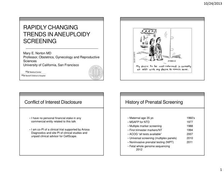

10/24/2013 RAPIDLY CHANGING TRENDS IN ANEUPLOIDY SCREENING Mary E. Norton MD Professor, Obstetrics, Gynecology and Reproductive Sciences University of California, San Francisco Conflict of Interest Disclosure History of Prenatal Screening • Maternal age 35 yo 1960’s • I have no personal financial stake in any commercial entity related to this talk • MSAFP for NTD 1977 • Multiple marker screening 1988 • I am co-PI of a clinical trial supported by Ariosa • First trimester markers/NT 1994 Diagnostics and site PI of clinical studies and • ACOG “all tests available” 2007 unpaid clinical advisor for CellScape. • Universal screening (multiplex panels) 2010 • Noninvasive prenatal testing (NIPT) 2011 • Fetal whole genome sequencing 2012 1
10/24/2013 Detection Rate of Prenatal Screening for Multiple marker screening Down syndrome has improved over time 120 • Uses a combination of first and second trimester serum 100 Detection Rate (%) analytes and ultrasound markers 80 60 • Primary purpose is screening for Down syndrome and 40 neural tube defects 20 0 • Can also detect other birth defects and abnormalities • In absence of fetal anomalies, abnormal levels of some analytes are associated with adverse obstetric outcomes Fetal Anomalies Associated with Fetal Anomalies Associated with Elevated MSAFP Elevated MSAFP • Open fetal anomalies • Open fetal anomalies • Neural tube defects (spina bifida/anencephaly) • Neural tube defects (spina bifida/anencephaly) Ventral wall defects (omphalocele/gastroschisis) • Ventral wall defects (omphalocele/gastroschisis) Chromosome abnormalities • Chromosome abnormalities Triploidy • Triploidy Severe renal abnormalities • Severe renal abnormalities Bilateral renal agenesis • Bilateral renal agenesis Autosomal recessive polycystic kidney disease • Autosomal recessive polycystic kidney disease Congenital skin disorders • Congenital skin disorders Upper GI tract obstruction • Upper GI tract obstruction 2
10/24/2013 Analyte Patterns Nuchal translucency • Increased NT highly associated with aneuploidy • Also with several other abnormalities UpToDate, 2008 Genetic Disorders Detected In Fetuses With Nonchromosomal Defects and Increased Enlarged Nuchal Translucency Nuchal Translucency Euploid NT (mm) Genetic disorders and fetuses (n) neurodevelopmental • Major cardiac defects delay • Diaphragmatic hernia Mangione et al., 2001 202 > 3mm 1/202 (0.5%) • Omphalocele Souka et al., 2001 1320 > 3.5 mm 44/1320 (3.3%) • Limb body wall defect • Fetal akinesia Senat et al., 2002 89 > 4 mm 4/62 (6.4%) • Noonan syndrome > 95 th % Bilardo et al., 2007 425 23/425 (5.4%) • Skeletal dysplasias • Other structural and genetic disorders Total 2271 72/1629 (4.4%) (range 0.5-6.4%) Bilardo Prenatal Diagnosis 2010 3
10/24/2013 Abnormal Analytes and 3d Trimester Fetal cardiac defects and enlarged NT Pregnancy Complications NT Incidence CHD • Several patterns of abnormal analytes have been associated with placental dysfunction <2.5mm 0.16% • Low PAPP-A, low uE3, high AFP, hCG, inhibin 2.4-3.4mm 1% • 3d trimester pregnancy complications 3.5-4.5mm 3% • Preeclampsia, early fetal loss, late fetal loss, 4.5-5.4mm 7% preterm delivery, IUGR 5.4-6.4mm 20% • Risk of adverse outcomes increases with higher levels >6.5mm 30% or with multiple abnormal analytes Souka, 2005 Odds Ratios for Outcomes Associated Association between Adverse Outcomes with Abnormal Analytes and Serum Analytes Analyte IUFD PTB IUGR PreE PAPP-A <0.29 MoM 3.0 3.3 4.64 1.79 PAPP-A <0.42 MoM 2.15 1.9 2.81 1.54 hCG >2.0 MoM 1.5-4.7 1.7-2.8 1.8-4.8 2.4 AFP >2.5 MoM 4.4-9.8 1.8-4.8 1.6-4.0 3.8 Inh A >2.0 MoM 2.4 2.4 2.4 2.4 uE3 <0.5 MoM 3.3 2.3 Dugoff, Obstet Gynecol, 2010 4
10/24/2013 Is detection of abnormal analytes helpful? Is detection of abnormal analytes helpful? Bujold et al, 2010, Obstet Gynecol Roberge et al, Ultrasound Obstet Gynecol 2013 Meta-analysis of studies of aspirin in high risk women Meta-analysis of studies of aspirin in high risk women Disorder ASA No ASA RR (CI) Disorder ASA No ASA RR (CI) Perinatal death 1.1% 4.0% 0.41 (0.19-0.92) PreE 9.3% 21.3% 0.47 (0.34-.65) Preeclampsia 7.6% 17.9% 0.47 (0.36-0.62) Severe PreE 7% 15% 0.09 (0.02-.37) Severe PreE 1.5% 12.3% 0.18 (0.08-0.41) IUGR 7% 16.3% 0.44 (0.30-.65) IUGR 8.0% 17.6% 0.46 (0.33-0.64) Preterm birth 4.8% 13.4% 0.35 (0.22-0.57) ASA decreased risks only when started at <16 weeks gestation ASA decreased risks only when started at <16 weeks gestation Noninvasive Prenatal Testing (NIPT) Analysis of cell free DNA using Cell Free DNA • Tests for aneuploidy by directly sequencing of fetal DNA, largely derived from placenta • Compared with current screening which uses indirect measurements of protein products • NIPT for detection of trisomy 21 has greater sensitivity and much greater specificity than multiple marker screening Zhong, X, Holzgreve, W, Glob. libr. women's med 2009 5
10/24/2013 Non-invasive Prenatal Testing (NIPT) Non-invasive Prenatal Testing (NIPT) vs First Trimester Screening (FTS) vs First Trimester Screening (FTS) Disorder Test Detection rate False positive rate Disorder Test Detection rate False positive rate Trisomy 21 NIPT 99.4% 0.15% Trisomy 21 NIPT 99.4% 0.15% FTS 94% 5.8% FTS 94% 5.8% Trisomy 18 NIPT 98.5% 0.2% Trisomy 18 NIPT 98.5% 0.2% FTS 100% 0.3% FTS 100% 0.3% Trisomy 13 NIPT 86% 0.7% Trisomy 13 NIPT 86% 0.7% FTS 100% 0.3% FTS 100% 0.3% Alamillo et al. 2013 False positive rate of NIPT Placental mosaicism and T13 and 18 As you add additional tests, the false positive rate • Survival rates of T13, 18 and 21 are relatively low, but increases vary • Study of karyotype of cytotrophoblast, villous stroma, chorion, amnion, and cord blood w/T13 and T18 infants T21 0.1% % cells trisomic in % cells trisomic in Fetal T18 0.5% trisomy cytotrophoblast all other tissues T13 0.7% T13, T18 30% (average) 100% 45X ~1.0% (n=14) Other SCA ? 100% 100% T21 (n=12) Total >2.3% NIPT false negatives and positives more likely in T13 and T18 due to underlying biology of fetal and placental development Kalousek et al, 1989 6
10/24/2013 Natural X Chromosome Loss NIPT is more precise for T21, T18 • 665 women (0-80yrs) • Lymphocyte cultures on 19,650 cells • G-banding analysis for presence of 1 or 2 X chromosomes Russell et al, 2007 NIPT Current NT + serum screen NIPT is more precise for T21, T18 NIPT is more precise for T21, T18 Other abnormalities Other abnormalities FTS NIPT 8/8 T21 8/8 T21 NIPT Current NT + serum screen 3/3 T18 2/3 T18; 1/3 no result 7/7 others (45X; triploidy; deletions and duplications) Nicolaides et al, 2012 7
10/24/2013 “Nearly a third of abnormalities found after NIPT is more precise for T21, T18 first trimester screening are different than expected” Alamillo et al. 2013 Other abnormalities NIPT FTS • N=23,329 cases of FTS over 10 years • 6.3% screen positive • 5.7% for T21; 0.4% for T13/18; 0.3% for both • 97 had a chromosome abnormality (1/240) 55% 100% (1/500) • 47 Down syndrome (T21) (10/18) (18/18) • 22 Trisomy 13 or 18 8/8 T21 • 29 Other chromosome abnormalities 8/8 T21 3/3 T18 • Range of severity from mild to lethal 2/3 T18; 1/3 no result 7/7 others (45X; triploidy; • Detected by combination of NT and analytes deletions and duplications) Nicolaides et al, 2012 DS and T18 make up 2/3 of aneuploidies Disorders potentially detectable by serum screening and NIPT detectable by karyotype NIPT Current Screening • Trisomy 21 • Trisomy 21 • Trisomy 18 • Trisomy 13 • Trisomy 18 • Some sex chromosomes • Trisomy 13 • Triploidy • Some sex • Other rare aneuploidies chromosomes • Congenital heart defects • Noonan syndrome • Neural tube defects • Ventral wall defects • Congenital adrenal hypoplasia • Smith Lemli Opitz syndrome • Steroid sulfatase deficiency • Poor OB outcomes (IUGR, PreE, PTB) 8
10/24/2013 Chromosomal Microarray (CMA) for Rate of aneuploidy varies by maternal age Prenatal Diagnosis • >35 yo 43% of abnormalities will be missed by NIPT • <35 yo 75% of abnormalities will be missed by NIPT • This includes only those that are detectable by karyotype • Rate is significantly higher if include those detectable by chromosomal microarray ACMG , Statement on Noninvasive Prenatal Screening, 2013 Diagnostic Yield in Cases with Normal Abnormalities detectable per 1000 births Karyotype Indication for Clinically Relevant 45 Testing (N=96) 40 U/S Anomaly 35 6.0% 30 NIPT N=755 AMA 25 1.7% Amnio/karyotype 20 N=1,966 15 Amnio/microarra Positive Screen 1.7% y 10 N=729 5 Other 0 1.3% 20 yo 25yo 30yo 35yo 40yo N=372 Wapner et al 2012; Hook 1983 9
Recommend
More recommend