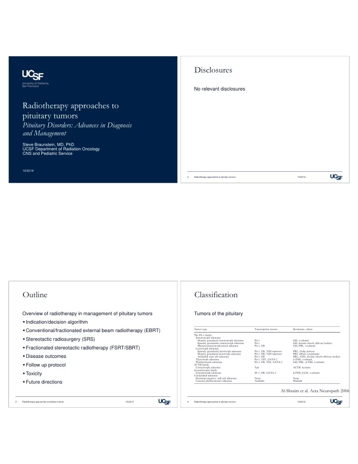

Disclosures No relevant disclosures Radiotherapy approaches to pituitary tumors Pituitary Disorders: Advances in Diagnosis and Management Steve Braunstein, MD, PhD UCSF Department of Radiation Oncology CNS and Pediatric Service 10/22/16 2 Radiotherapy approaches to pituitary tumors 10/22/16 Outline Classification Overview of radiotherapy in management of pituitary tumors Tumors of the pituitary � Indication/decision algorithm � Conventional/fractionated external beam radiotherapy (EBRT) Tumor type Transcription factors Hormones, others The Pit-1 family � Stereotactic radiosurgery (SRS) Somatotroph adenoma Densely granulated somatotroph adenoma Pit-1 GH, a -subunit Sparsely granulated somatotroph adenoma Pit-1 GH, keratin whorls (fibrous bodies) Mammosomatotroph/mixed adenoma Pit-1, ER GH, PRL, a -subunit � Fractionated stereotactic radiotherapy (FSRT/SBRT) Lactotroph adenoma Sparsely granulated lactotroph adenoma Pit-1, ER, ?GH-repressor PRL, Golgi pattern Densely granulated lactotroph adenoma Pit-1, ER, ?GH-repressor PRL diffuse cytoplasmic � Disease outcomes Acidophil stem cell adenoma Pit-1, ER PRL, (GH), keratin whorls (fibrous bodies) Thyrotroph adenoma Pit-1, TEF, GATA-2 b -TSH, a -subunit Plurihormonal adenoma Pit-1, ER, TEF, GATA-2 GH, PRL, b -TSH, a -subunit � Follow up protocol ACTH family Corticotroph adenoma Tpit ACTH, keratins Gonadotropin family � Toxicity Gonadotroph adenoma SF-1, ER, GATA-2 b -FSH, b -LH, a -subunit Unclassified adenoma Hormone-negative/ null cell adenoma None None � Future directions Unusual plurihormonal adenoma ?multiple Multiple Al-Shraim et al. Acta Neuropath 2006 3 Radiotherapy approaches to pituitary tumors 10/22/16 4 Radiotherapy approaches to pituitary tumors 10/22/16
Classification Post surgical outcomes Tumors of the pituitary Overall local control is 50-80% following resection � Pituitary adenoma Recurrence Risk for non-functional tumors: • Microadenoma (<1cm) Post-op MRI 5 yr 10 yr GTR 10-20% 30% • Macroadenoma ( ≥ 1cm) STR 25-40% >50% • Functional • Non-functional � Pituitary carcinoma � Metastases (breast and lung) Cortet-Rudelli et al. Annales d’Endocrinologie 2015 5 Radiotherapy approaches to pituitary tumors 10/22/16 6 Radiotherapy approaches to pituitary tumors 10/22/16 Radiotherapy indications Radiotherapy approach Surgical local control 50-80% Pre-treatment workup � Medically inoperable (panhypopituitarism) � Complete endocrine evaluation � Subtotal resection (persistent hypersecretion) � Visual field testing � Large tumor with extrasellar extension Cessation of suppressive medications � Recurrence � Non-randomized data � Pituitary carcinoma (high mitotic index, invasive features) 7 Radiotherapy approaches to pituitary tumors 10/22/16 8 Radiotherapy approaches to pituitary tumors 10/22/16
Radiotherapy approach Fractionated external beam radiotherapy Effect of endocrine suppression Conventionally/classically fractionated (LINAC-based) � Nonfunctioning: 45- 50 Gy � Functioning: 50.4-54 Gy HS No-HS Sheehan et al. JNS 2011 9 Radiotherapy approaches to pituitary tumors 10/22/16 10 Radiotherapy approaches to pituitary tumors 10/22/16 Fractionated external beam radiotherapy Fractionated external beam radiotherapy Conventionally/classically fractionated (LINAC-based) 45 Gy McCoullough WM IJROBP 1991, 95% LC Preferred when large pituitary adenomas and/or when lesion is < 2mm from optic chiasm Zeirhut et al. IJROBP 1995 11 Radiotherapy approaches to pituitary tumors 10/22/16 12 Radiotherapy approaches to pituitary tumors 10/22/16
Stereotactic radiosurgery Stereotactic radiosurgery Single session radiosurgery (Gamma knife, Cyberknife, LINAC) Single session radiosurgery (Gamma knife, Cyberknife, LINAC) � Nonfunctioning: 12-20 Gy � Functioning: 15-30 Gy Preferred to decrease dose to hypothalamus and cortical brain 13 Radiotherapy approaches to pituitary tumors 10/22/16 14 Radiotherapy approaches to pituitary tumors 10/22/16 Fractionated stereotactic radiotherapy Outcomes Multisession radiosurgery (Gamma knife, Cyberknife, LINAC) � Nonfunctioning: 25-30 Gy in 5 fractions Tumor type Tteatment protocol Disease free survival 10 yr � Functioning: 30-35 Gy in 5 fractions Non-functioning Surgery � obs vs RT 90% RT alone 80% GH-secreting Surgery � obs vs RT 70-80% RT alone 60-70% Prolactin secreting Obs vs MM vs Sg vs RT 80-90% Preferred when pituitary lesion is > 3cm and/or ACTH-secreting Surgery � obs vs RT 50-60% (more rapid) when lesion is < 2mm from optic chiasm RT alone 50-60% TSH-sectreing Surgery � RT 40-50% 15 Radiotherapy approaches to pituitary tumors 10/22/16 16 Radiotherapy approaches to pituitary tumors 10/22/16
Outcomes Outcomes Functional control Overall survival � ACTH normalizes < 1yr � No difference in OS among: � Prolactin > 1yr • Surgery � Growth hormone > 1yr (50% at 2 yr, 70% at 10 yr) • Surgery + RT • RT alone � Choose therapy based on minimizing side effects 17 Radiotherapy approaches to pituitary tumors 10/22/16 18 Radiotherapy approaches to pituitary tumors 10/22/16 Follow up Outcomes Delayed response and toxicity Pituitary function � MRI 6 and 12 months, then annually � Endocrine evaluation every 6-12 months � Formal visual field testing annually Xu et al. Neurosurgery 2013 19 Radiotherapy approaches to pituitary tumors 10/22/16 20 Radiotherapy approaches to pituitary tumors 10/22/16
Outcomes Outcomes/Toxicity Visual function � EBRT: 50-54 Gy Secondary Malignancy � SRS: 8 Gy 70 Applicability of models to predict RION from conventional to SRS fractionations Pendulum Two opposed Models and literature indicate Model: LQ extrapolation from 1.8 Gy/fx, 59.4 Gy with α / β =3.3 60 better tolerance at lower dose treatment lateral fields Three fields Total Model: LQ extrapolation from 1.8 Gy/fx, 59.4 Gy with α / β =1.6 per fraction. Model: Iso Neuret(NSD) = 60 Gy, 1.8 Gy/fx 50 Number of patients 38 138 61 237 Total Dose (Gy) Model: Iso Optic RET = 8.9 Gy Median age/years at radiotherapy (range) 54 (24–75) 55 (18–79) 62 (16–82) 56 (16–82) Literature Findings: > 10% Incidence RION Number of person-years at risk 750 1861 499 3110 40 Literature Findings: 1-9% Incidence RION Number of second primary tumours within area of radiotherapy 1 4 0 5 Literature Findings: No Incidence RION Total number of second primary tumours 5 20 5 30 30 Number of tumours/10 3 person-years at risk 6·6 10·7 10·0 9·6 Majority of published data pre-date planning and 20 treatment delivery technology that allows for steep dose gradients in or near optic 10 structures. Effect on partial Lack of published data in volume tolerance needs Only a few detailed publications in SRS region hypo-fractionation region further exploration. 0 0 2 4 6 8 10 12 14 Norberg et al. Clin Endocrinology 2008 Mayo et al. IJROBP 2010 Dose per Fraction (Gy) 21 Radiotherapy approaches to pituitary tumors 10/22/16 22 Radiotherapy approaches to pituitary tumors 10/22/16 Outcomes/Toxicity Pituitary carcinoma Secondary Malignancy Sex/age (in years) Years between radiotherapy at radiotherapy for and the diagnosis of a Number of fields used Type of tumour pituitary adenoma second primary tumour and radiation dose received (in Gy) Glioma (astrocytoma grade III) Male/55 7 Two opposed lateral fields, 40 Meningioma Male/46 9 Two opposed lateral fields, 45 Meningioma Male/54 First treatment 24 First treatment: pendulum, 41 Second treatment 1 Second treatment: two opposed lateral fields, 31 Cancer in the parotid gland Female/73 8 Two opposed lateral fields, 42 Squamous cell carcinoma in the external ear Male/51 9 Two opposed lateral fields, 42 Norberg et al. Clin Endocrinology 2008 23 Radiotherapy approaches to pituitary tumors 10/22/16 24 Radiotherapy approaches to pituitary tumors 10/22/16
Pituitary carcinoma Future directions � Proton � Heavy particle � Adaptive hybrid radiosurgery � LGKS � Molecular prognostic markers/indices Heaney J Clin Endocrinol Metab 2011 25 Radiotherapy approaches to pituitary tumors 10/22/16 26 Radiotherapy approaches to pituitary tumors 10/22/16 Future directions Molecular Prognostic indices Ki-67, p53, MI Diagnostic Parameters Cut-off Sensitivity Specitifity Youden-Index Accuracy in % AUC 95 % CI OR 95 % CI P -value for APAs/TPAs of AUC of OR Ki-67 pos. nuclei in % ≥ 4 0.95 0.97 0.92 96 0.98 [0.96; 1.0] 5.2 [3.43; 7.83] <0.001 P53 pos. nuclei in % ≥ 2 0.85 0.93 0.78 90 0.94 [0.90; 0.97] 3.1 [2.31; 4.04] <0.001 Mitotic Index in 10 HPF ≥ 2 0.90 0.74 0.64 79 0.89 [0.84; 0.93] 2.1 [1.70; 2.57] <0.001 Invasiveness Yes 0.88 0.53 0.41 64 - - 8.2 [3.66; 18.42] <0.001 The proposed threshold values for Ki-67, p53, number of mitotic figures in 10 HPF (high power fields) and the status of invasive tumor growth, to distinguish APA and TPA are shown with their respective statistical values OR odds ratio, AUC area under curve, CI confidence interval Miermeister et al. Acta Neuropathologica Comm 2015 27 Presentation Title and/or Sub Brand Name Here 10/22/16
Recommend
More recommend