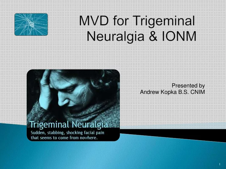

Presented by Andrew Kopka B.S. CNIM 1
2
Common EP’s / recordings used in the O.R. • SSEP - Somatosensory evoked potentials • TcMEP - Transcranial motor evoked potentials • BAER - Brainstem auditory evoked responses • EMG - Free Run • EMG -Triggered • EEG - Electroencephalography 3
Cranial Nerve V: • Mixed nerve • Largest cranial nerve • 2 roots from venterolateral of the pons • Large sensory root (Portio major) and small motor root (Portio minor) 4
• Cranial Nerve V: • Major sensory face • Motor for mastication • 3 divisions Ophthalmic division V1 Maxillary division V2 Mandibular division V3 Temporalis Masseter 5
V1 - Ophthalmic Division 6
V2 - Maxillary Division 7
V3 - Mandibular Division 8
• Trigeminal Neuralgia (TN) is neuropathic facial pain arising from the trigeminal nerve. • The pain is intense, sharp, electric shock-like pain in the face, lasting periods seconds, minutes, hours. • Incidence 4-5 cases : 100.000 • TN or Tic Douloureux occur patients > 45 years. • Male : Female ratio 1 : 1.5 • Unilateral (97%). Most affected V2 and V3. 9
• Light pressure at “trigger points” can trigger attacks. • Unpredictable symptom free intervals. • Patient biggest ambitions • Eating, shaving, applying makeup……… 10
Classification Trigeminal Neuralgia TYPICAL TN PRE-TN ATYPICAL TN 7 TYPES MULTIPLE POST- SCLEROSIS TRAUMIC RELATED TN TN 2ndary FAILED TN TN 11
1. Most common form. Severe sudden excruciating unilateral pain face. 2. Intense, stabbing, electrical shock-like pain. 3. Blood vessels compressing the trigeminal nerve root at the REZ - trigeminal nerve enters brain stem Superior cerebellar artery (SCA) Anterior inferior cerebellar artery (AICA) 4. Repeated vascular pulsations causes demyelination & injury to the trigeminal nerve - hyperactivity trigeminal nucleus - in TN pain 5. Frequently pain free between attacks. 6. Lasting only seconds - minutes - hours. 7. Each attack spontaneous or be triggered by specific stimulation. 8. Common triggers include touch, talking, eating, drinking, chewing, tooth brushing, hair combing 12
(c) (a) No compression CN V Vascular compression present 13
Typical TN Progression 14
• Clinical history • Clinical examination • CT scan and MRI • MRA 15
Medication: Anticonvulsants • Carbamazepine (Tegretol) • Drug of choice for TN, effective dose 600 -1200 mg/ day for 3-4 x/ day • Side effect: drowsiness, mental confusion, dizziness, ataxia • Oxycarbazepine (Trileptal ) Side effect: nausea, fatigue, tremor, anemia…….. • • Dose : 2 x 300mg, maximum dose : 2400-3000 mg/day • Phenytoin (Dilantin) Dose: 300-500mg/day for 3x day • • Side effects: Nystagmus, dysarthria, gingival hyperplasia, hypertrichosis, allergic skin rash • Gabapentin (Neurontin) • Dose: 300-1200mg/day • Side effects: drowsiness, ataxia, fatigue 16
• “It’s the surgeons decision”…… • General use EP’s and recordings for MVD’s: • * BAER’s SSEP’s • • *Triggered and Free run EMG 17
• BAER’s reflect the neurological responses of the 8 th cranial nerve (vestibulocochlear nerve), following activation at the cochlea via a click stimulus, to various generator sites along the 8 th cranial nerve and the brainstem. The first five waves are resistant to anesthesia and therefore are well suited to IONM. The multiple generator sites allow relative localization of insults during surgeries involving the brainstem and the 8 th cranial nerve pathways. Goal! - early warning impending neurological hearing deficits! 18
• BAER’s are elicited by delivering a click stimulus to the ear. To avoid contributions from the contralateral ear, masking white noise is delivered to the contralateral ear at approx. 40dB nHL. • Many averaged trials are required to record reliable responses. Recommended stimulation rates are between 5- 15 Hz (that’s 5 -15 stimuli/sec) with 11 Hz being a reasonable balance. Recommended stimulation and recording parameters are as follows: High Am Am Typical Stim. Stim Stim. Low Freq Freq p latencies es Intensity sity Durati tion on Rate Filter r (Hz) Filter r (μV) (ms) (dB) (ms) (Hz) (Hz) 1500- 0.3- 75-110 BAER 30-100 1.5-10 0.1 5-15 3000 3 dB 19
• BAER pathways : Wave I - Cochlea & extracranial cochlear nerve (CN VIII) Wave II - Intracranial portion acoustic nerve & cochlear nucleus (Medulla) Wave III - Superior olivary complex (Pons) Wave IV - Lateral lemniscus (Pons) Wave V- Inferior colliculus (Midbrain) V I IV II III 20
Important* Contralateral ear and 2 -3 channels used to record • • Cv2 - Cz generally have poorly BAER’s following the International defined Waves I-IV, but have a well 10-20 System: defined Wave V. Channel 1: A1 - Cz • Note* Ipsilateral ear to contralateral • Channel 2: A2 - Cz • ear may also be used (A1-A2) Channel 3: Cv2 (inion) - Cz Mastoid substitute: A1 and A2 • • 21
There are 5 principle features used to assess routine BAER’s 1. I-V interpeak interval 4. V/I amplitude ratio 2. I-III interpeak interval 5. Presence of wave I-V 3. III-V interval V IV III I II 22
Point change significant?........institutional - baseline responses - a latency increase wave V of more than 1.0ms from baseline - a amplitude reduction of 50% from baseline Changes I-III interpeak latency (IPL): • Suggests disturbance along the eighth nerve close to the cochlea and the lower pons/cochlear nucleus. Often due to stretching/manipulation 8 th nerve Changes III-V IPL: • Suggest disturbance between the lower pons/superior olivary complex and the midbrain/inferior colliculus. Often due to cerebellar compression due to retractor placement, or hypotension Changes wave V latency: • Gradual latency/amp changes can begin w/ CPA exposure. Due to variety factors: stretch 8 th CN, retractor placement, or cold irrigation . Abrupt loss wave I: • Loss of wave 1, w / wo loss waves II - V due to compression/stretching auditory artery (labyrinthine artery) results – ischemia cochlea. Rapid: persists 15min = perm hearing loss 23
Free run EMG: • Free run electromyography (EMG) records the patterns electrical activity assoc. w/ skeletal muscles: continuous, live real-time. Since specific muscles are attached to specific nerves, nerve function can be implied from the type of activity seen in the EMG recording. • Recorded subdermal electrodes: corresponding muscles assoc. neuro structures monitored. • Resting muscle/assoc. nerve are electrically silent. When the nerve is irritated or injured, it will fire spontaneously, causing reciprocal firing in the muscle. This manifests as muscle responses “firing” occur in several patterns indicating degrees of irritation or injury including: 24
spikes (individual discharges) bursts (brief flurries of discharges) 25
train activity (more persistent regularly repeating discharge patterns) neurotonic discharges (persistent prolonged bursting) 26
Triggered EMG: • Neuro. structure/nerve stimulated and a CMAP is recorded in corresponding muscles innervated by the neurological structure. CN 7 • Used to: • ID nervous tissue • ID neurological structures (cranial nerve, nerve root) • Integrity nerve (damage) CN 5 27
• Evaluate the health/state of muscles and motor neurons controlling muscles • Recording parameters EMG: Low High Gain/ Typical Stim. Stim Stim. Time Freq Freq sensiti sitivit vit latencie Intensit nsit Durati tio Rate Base Filter Filter y s s (ms) y y (mA) n ( (ms) (Hz) (ms) (Hz) (Hz) (μV) Free ee 1500- 30-100 20-50/div n/a n/a n/a n/a 100/div EMG 3000 1500- Trig 30-100 20-50/div 10-25 10-25 0.2 1-4 1-10/div EMG 3000 • MVD: Needle electrode placement • Masseter and Temporalis (CN V) • Obic. oculi and Obic. Oris (CN VII)……….reasons? • Trapezius (CN XI) control 28
Teflon pad 29
http://www.youtube.com/watch?v=FDQa95DqHes 30
• Trigeminal nerve (CN V) separate into ophthalmic (V1), maxillary (V2), and mandibular (V3) branches provided major sensory feedback for the face and motor innervation masseter and temporalis muscles. • Trigeminal Neuralgia (TN) is shock-like neuropathic facial pain arising from compression trigeminal nerve via SCA (AICA). • Treatment for typical TN….. Medication: if pharmacologic treatment fails….. surgical procedure. • MVD: craniotomy decompresses CN V by placing Teflon pads b/w SCA (or AICA) and CN V. • BAER’s and Trig / Free run EMG provide intraoperative feedback as to the state of CN V, CN VII, and CN VIII helping to protect these neuro structures during surgical procedure and preserve hearing, sensation to the face, and motor control of the facial muscles. 31
Recommend
More recommend