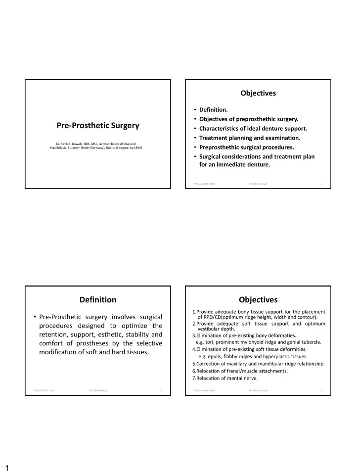

Objectives • Definition. • Objectives of preprosthethic surgery. Pre-Prosthetic Surgery • Characteristics of ideal denture support. • Treatment planning and examination. Dr. Rafik Al Kowafi BDS, MSc, German board of Oral and • Preprosthethic surgical procedures. Maxillofacial Surgery ( Berlin-Germany), Doctoral degree by LBMS • Surgical considerations and treatment plan for an immediate denture. 3 March 2016 LIMU Dr. Rafik Al Kowafi 2 Definition Objectives 1.Provide adequate bony tissue support for the placement • Pre-Prosthetic surgery involves surgical of RPD/CD(optimum ridge height, width and contour). 2.Provide adequate soft tissue support and optimum procedures designed to optimize the vestibular depth. retention, support, esthetic, stability and 3.Elimination of pre-existing bony deformaties. e.g. tori, prominent mylohyoid ridge and genial tubercle. comfort of prostheses by the selective 4.Elimination of pre-existing soft tissue deformities. modification of soft and hard tissues. e.g. epulis, flabby ridges and hyperplastic tissues. 5.Correction of maxillary and mandibular ridge relationship. 6.Relocation of frenal/muscle attachments. 7.Relocation of mental nerve. 3 March 2016 LIMU Dr. Rafik Al Kowafi 3 3 March 2016 LIMU Dr. Rafik Al Kowafi 4 1
Characteristics of ideal denture Characteristics of ideal denture support support 1. No evidence of intraoral or extraoral pathologic 6. Proper posterior tuberosity notching . conditions. 7. Adequate attached keratinized mucosa in the primary 2. Proper inter-arch jaw relationship in anteroposterior, denture-bearing area. transverse, and vertical dimensions. 8. Adequate vestibular depth for prosthesis extension. 3. Alveolar processes that are as large as possible and of 9. Added strength where mandibular fracture may occur. proper configuration (the ideal shape of the alveolar 10. Protection of the neurovascular bundle. process is a broad U-shaped ridge, with the vertical 11. Adequate bony support and attached soft tissue components as parallel as possible) covering to facilitate implant placement when 4. No bony or soft tissue protuberances or undercuts. necessary. 5. Adequate palatal vault form. 3 March 2016 LIMU Dr. Rafik Al Kowafi 5 3 March 2016 LIMU Dr. Rafik Al Kowafi 6 Treatment planning and examination I. Extraoral examination: (Facial esthetic examination) I. Presence of unsupported upper lip. II. Poor vermilion show. III. Loss of nasolabial fold or decreased nasolabial fold. IV. Poor/obtuse nasolabial angle with poor projection. V. Excessive lower lip show. A , Ideal shape of alveolar process in denture-bearing area. B-E , Diagrammatic representation of progression of bone resorption in mandible after tooth extraction. 3 March 2016 LIMU Dr. Rafik Al Kowafi 7 3 March 2016 LIMU Dr. Rafik Al Kowafi 8 2
Treatment planning and examination Treatment planning and examination II. Intraoral examination (hard and soft tissues): 6. Interarch relationship. 7. Adequate post-tuberosity notching. 1. Ridge form and contour: a. Height and width of the ridge. 8. The amount of keratinized tissue and poorly keratinized or freely movable tissue. b. Quality of the ridge-whether flabby, mobile tissue is present over the ridge. 9. Inflammatory areas, scars, ulcers, hyperplastic c. Presence of any gross irregularities in the ridge. tissues due to ill-fitting dentures should be looked 2. Presence of any exostosis, undercuts, prominences, tori, for. sharp mylohyoid ridge or severe resorption of external 10. Frenal attachments in relation to the alveolar crest. oblique ridge. 11. On the lingual aspect, mylohyoid muscle attachmen 3. Buccal and labial, as well as lingual vestibules evaluation for and genioglossus muscle attachment should be depth and type of soft tissue. checked. 4. Examination of palatal vault. 12. Tongue size and movement is also important for the 5. Tuberosity area - undercuts, hyperplastic tissue, flabby ridge, stability of the denture. etc. Height, width, fibrous or excess bony tuberosity can impair the arch space for fabrication of full or partial denture. 3 March 2016 LIMU Dr. Rafik Al Kowafi 9 3 March 2016 LIMU Dr. Rafik Al Kowafi 10 Preprosthethic surgical procedures Treatment planning and examination A. Alveolar Ridge Correction: I. Recontouring of alveolar ridges: III. Radiographic 1. Simple Alveoloplasty Associated with Removal of Multiple Teeth examination: 2. Intraseptal Alveoloplasty 3. Maxillary Tuberosity Reduction 1. OPG. 4. Buccal Exostosis and Excessive Undercuts 5. Lateral Palatal Exostosis 2. Lateral cephalometric 6. Mylohyoid Ridge Reduction radiograph. 7. Genial Tubercle Reduction 3. Computed tomography: II. TORI REMOVAL: 1. Maxillary Tori . - Dental CT scan. 2. Mandibular Tori . - 3D CT-Scan. III. Soft tissue abnormalities: 1. Maxillary Tuberosity Reduction (Soft Tissue) - CBCT. 2. Mandibular Retromolar Pad Reduction IV. Diagnostic models: 3. Lateral Palatal Soft Tissue Excess 4. Unsupported Hypermobile Tissue Mounted on articulator 5. Inflammatory Fibrous Hyperplasia 6. Inflammatory Papillary Hyperplasia of the Palate with proper vertical 7. Labial Frenectomy dimension. 8. Lingual Frenectomy B. Alveolar ridge augmentation. C. Alveolar ridge extension (vestibuloplasty). D. Alveolar ridge distraction. 3 March 2016 LIMU Dr. Rafik Al Kowafi 11 3 March 2016 LIMU Dr. Rafik Al Kowafi 12 3
A- Alveolar Ridge Correction 1- Simple Alveoloplasty 1- Simple Alveoloplasty • Alveoloplasty refers to the surgical recontouring of the alveolar process. • It can be primary and secondary alveoloplasty. – Primary alveoloplasty is always done at the time of single or multiple extractions. – Secondary alvoplasty is done on the edentulous ridge late after the extraction. • This procedure includes: a. Extraction of the tooth/teeth. b. Incision and reflection of the gingivae. c. Smoothing of alveolar bone by using bone File, rongeur forceps or bone Bur. d. Care of wound. a Periapical radiograph of the region of the canine and first premolar of themandible. b Clinical photograph. e. Suturing of the mucoperiosteum. Supraeruption of teeth and a high alveolar ridge are noted 3 March 2016 LIMU Dr. Rafik Al Kowafi 13 3 March 2016 LIMU Dr. Rafik Al Kowafi 14 1- Simple Alveoloplasty 1- Simple Alveoloplasty a, b. Removal of wedge-shaped portions of mucosa from the alveolar ridge, from the area mesial and distal to the a, b. Reflection of the mucoperiosteum and removal of bone margins of the wound with a rongeur sockets 3 March 2016 LIMU Dr. Rafik Al Kowafi 15 3 March 2016 LIMU Dr. Rafik Al Kowafi 16 4
1- Simple Alveoloplasty 1- Simple Alveoloplasty a, b. Smoothing of the bone surface with a bone bur. a Diagrammatic illustration. b Clinical photograph a, b. a Operation site after placement of sutures. b Postoperative clinical photograph 1month after the surgical procedure 3 March 2016 LIMU Dr. Rafik Al Kowafi 17 3 March 2016 LIMU Dr. Rafik Al Kowafi 18 2- Intraseptal Alveoloplasty 2- Intraseptal Alveoloplasty (Dean’s intraseptal alveoloplasty) (Dean’s intraseptal alveoloplasty) • In this technique, the interseptal bone is removed followed by reposition of the labial cortical bone. • This technique is used in areas of adequate bone height, but have undercut at the depth of the labial vestibule.. • This is done by reflecting mucoperiosteal flap by making buccal and lingual parallel incisions to remove the interdemntal papilla, which is chronically inflamed. • The buccal mucoperiosteum is reflected to the level of the junction of free and attached mucosa with minimum elevation of the lingual mucoperiosteum. The interseptal bone is removed using rongeur or large surgical burs under copious saline irrigation, then digital compression is used to push the labial cortical bone inward, and the flap is repositioned back again and sutured. • Continuous suture is used to stabilize the flap against the alveolar ridge and not only to approximate the mucosal margins. 3 March 2016 LIMU Dr. Rafik Al Kowafi 19 3 March 2016 LIMU Dr. Rafik Al Kowafi 20 5
3- Maxillary Tuberosity Reduction 3- Maxillary Tuberosity Reduction • Enlarged tuberosity may involve either bone, soft tissues or both. Pre-operative X-ray is mandatory to rule out any pathological process (impacted teeth or fibrous dysplasia). • A crestal incision is made behind the tuberosity (using blade No. 12) then the excess bone is removed using rongeur or surgical bur under copious saline irrigation. The area is smoothened with the bone file, and a wedge of the soft tissue is excised to allow proper soft tissue closure. • The wound is closed with interrupted or continuous sutures. • The most common complication of this procedure is opening of the maxillary sinus. If there is small opening, soft tissue coverage can be achieved easily as long as there is no infection. 3 March 2016 LIMU Dr. Rafik Al Kowafi 21 3 March 2016 LIMU Dr. Rafik Al Kowafi 22 3- Maxillary Tuberosity Reduction 3- Maxillary Tuberosity Reduction 3 March 2016 LIMU Dr. Rafik Al Kowafi 23 3 March 2016 LIMU Dr. Rafik Al Kowafi 24 6
Recommend
More recommend