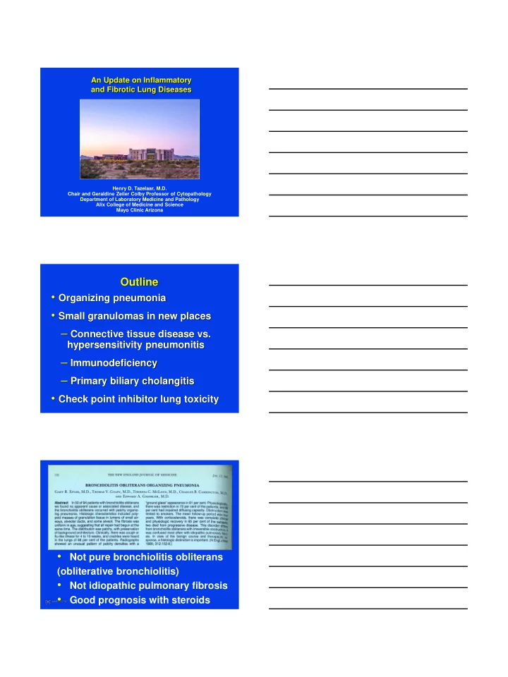

An Update on Inflammatory and Fibrotic Lung Diseases Henry D. Tazelaar, M.D. Chair and Geraldine Zeiler Colby Professor of Cytopathology Department of Laboratory Medicine and Pathology Alix College of Medicine and Science Mayo Clinic Arizona Outline • Organizing pneumonia • Small granulomas in new places – Connective tissue disease vs. hypersensitivity pneumonitis – Immunodeficiency – Primary biliary cholangitis • Check point inhibitor lung toxicity • Not pure bronchiolitis obliterans (obliterative bronchiolitis) • Not idiopathic pulmonary fibrosis • Good prognosis with steroids
ATS/ERS Classification Travis WD et al Am J Resp Crit Care Med 2002;165:277-304 Bronchiolitis obliterans a.k.a Constrictive bronchiolitis UIP
Organizing Pneumonia CP1060982-93
Bronchiolitis Obliterans with Organizing Pneumonia • Despite sometimes long history, changes were of “same age” • Some alveolar interstitial fibrosis-peribronchiolar • Honeycombing never seen Hyalinized Organizing Pneumonia trichrome Yousem SA et al Mod Pathol 1997;10:864-871
Hyalinized/Cicatricial/Fibrosing Organizing Pneumonia • 12 pts with cryptogenic disease • 55% had progressive or persistent CT infiltrates • 25% assoc. osseous metaplasia • Contribution of pre-existing non fibrotic lung ds, like emphysema which impairs healing? • Suggested poor steroid response Yousem SA Hum Pathol 2017; 64:76-82 Hyalinized/Cicatricial/Fibrosing Organizing Pneumonia • 10 pts identified by pattern • 20% assoc with radiologic ossification • Mimic of fibrotic NSIP • Non-progressive disease • Ehlers Danlos-1 pt Churg A et al Histopathol 2018; 72:846-854
Ehlers Danlos Syndrome Patient History • 41-year-old male real estate broker – CC: Cough and hemoptysis • Past Medical History – Asthma – Recurrent pneumothorax • Unilateral January 2016 • Bilateral October 2016 – Autoimmune serologies negative
4x 4x Ehlers-Danlos Syndrome Villefranche Prior Inheritance Genes classification 1 Nomenclature Pattern • Collagen defect COL5A1 and Classic Type I and II AD COL5A2 AD (likely) 2 Unknown 2 Hypermobility Type III AD 3 Vascular Type IV COL3A1 Kyphoscoliosis Type VI AR PLOD1 COL1A1 (VIIA) Arthrochalasia Type VIIA and B AD COL1A2 (VIIB) Dermatosparaxis Type VIIC AR ADAMTS2 Beighton P et al Am J Med Genet 1998;77(1):31-7
Differential Diagnosis for Hyalinizing OP • Ehlers Danlos syndrome • OP in NSIP • Fibroblast foci of UIP • Aspiration pneumonia with OP pattern and ossification
Hyalinized/Cicatricial/Fibrosing Organizing Pneumonia • New pattern to recognize • Prognostic significance unclear • Likely also occurs in association with other disease e.g. CTD • Other features may point to etiology-EDS, aspiration Churg A et al Histopathol 2018; 72:846-854 Q. A 58 year old woman with a history of Sjogren syndrome, gastroparesis, MALT lymphoma (treated with Rituximab and radiation) presented with a two day history of increasing shortness of breath. A CT about 1 month prior (done for cough and dyspnea) showed a stable infiltrate or scarring in the lingula … . a. Aspiration bronchiolitis b. CTD related changes c. Drug toxicity d. Hypersensitivity pneumonitis e. Non specific interstitial pneumonia
Q. A 58 year old woman with a history of Sjogren syndrome, gastroparesis MALT lymphoma (treated with Rituximab) presented with a two day history of increasing shortness of breath. A CT about 1 month prior (done for cough and dyspnea) showed a stable infiltrate or scarring in the lingula … . a. Aspiration bronchiolitis b. CTD related changes c. Drug toxicity d. Hypersensitivity pneumonitis e. Non specific interstitial pneumonia
Diagnosis Chronic bronchiolitis with features of follicular bronchiolitis with non-necrotizing granulomatous inflammation, and patchy mild cellular and fibrotic interstitial pneumonia, most consistent with underlying connective tissue disease Chronic Hypersensitivity Pn’itis (CHP, n=16) vs. Fibrotic Disease due to Connective Tissue Disease (CTD, n=12) • Reviewed 15 parameters • Germinal centers, prominent lymphoid aggregates and plasma cells favor CTD • Peribronchiolar metaplasia favors HP • Features that did not help: giant cells, granulomas, distribution of FiFo, pattern of fibrosis * Churg A et al Am J Surg Pathol 2017; 41:1403-1409 Peribronchiolar metasplasia
Chronic HP with UIP pattern Prominent peribronchiolar metaplasia
Favor Chronic HP Chronic Hypersensitivity Pn’it is (CHP) vs. Fibrotic Disease due to Connective Tissue Disease (CTD) • Challenging differential diagnosis • Other features - Favor CHP: air trapping on HRCT, identifiable antigen - Favor CTD: multi-compartment disease e.g. pleuritis, vasculopathy
Lung in Primary Biliary Cholangitis n=16, 94% women Feature Percent Lymphocytic inflammation 94 Mainly peribronchiolar Non necrotizing granulomas 81 UIP/NSIP patterns 52 Organizing pneumonia 44 Eosinophils 33 MALT lymphoma with light 6 chain deposition Lee HE et al Hum Pathol 2018;82:177-186 PBC PBC
Favor Chronic HP
Common Variable Immunodefiency (CVID) • Global immune dysfunction • B cells, T cells, cytokines – Explains combination of infectious, inflammatory, autoimmune, and neoplastic conditions – Significantly reduced serum IgG – Low serum IgA and / or IgM CVID Pulmonary Manifestations • Infection-pneumonia, bronchitis • Bronchiectasis • Asthma • Interstitial lung disease – Granulomatous-lymphocytic interstitial lung disease (GL-ILD)* – Organizing pneumonia *Bates CA et al J Allergy Clin Immunol 2004; 114: 415-21 So- called GL-ILD • Dyspnea • Restrictive PFT’s • HRCT: consolidation, ground-glass opacities, reticular opacities • Various histologies Bates CA et al J Allergy Clin Immunol 2004; 114: 415-21
So- called GL-ILD Histologic Features • Lymphocytic infiltrates/LIP • Non necrotizing granulomas (most, but not all) • Follicular bronchiolitis • Diffuse lymphoid hyperplasia • Prominent organizing pneumonia • Fibrosis (including honeycomb) Rao N et al Hum Pathol 2015;46:1306-1314
CD3 CD5 CD43 CD20
CD138 Kappa Lambda So- called GL-ILD Histologic Features • Wide spectrum of histologies • Possibly useful as a clinical term, but very confusing for pathologists! • Always need to exclude lymphoma • Granulomas don’t exclude lymphoma (20% of pulmonary MALT lymphomas) History • 76 yr. old M 10 pack yr smoker • Referred for possible bronchoscopy • History of metastatic melanoma • Started on Immunotherapy with Pembrolizumab 10 months ago
History Hospitalized • Weight loss of 6-8 lbs. over the last 4 weeks • Noted dyspnea on exertion for 4 weeks • Dry cough for 3 weeks • Low grade fever and chills for the past 10 days CT 3 Months Prior
Transbronchial biopsy
Pathologic Diagnosis? • Organizing Pneumonia – DDx: infection, drug reaction, connective tissue disease, aspiration, and as an idiopathic entity (cryptogenic organizing pneumonia) Final Clinical Diagnosis • BAL and special stains negative for infection • ? Pembrolizumab-induced organizing pneumonia • Pt started on 60 mg of prednisone • Improved over the next few weeks with tapering of steroids over 2 months Check Point Inhibitor- Assoc ILD, n=64 Radiologic Patterns % Organizing pneumonia (OP) 23.4 Hypersensitivity Pneumonia 15.6 NSIP plus OP 9.4 NSIP 7.8 Bronchiolitis 6.3 NSIP plus bronchiolitis 1.6 Not classified 35.9 Delaunay M et al. Eur Resp J 2017;50:1700050
Check Point Inhibitor- Pathology Patterns Diffuse alveolar damage Organizing pneumonia (OP) +/- fibrin Hypersensitivity pneumonitis/granulomatous pneumonitis NSIP-cellular interstitial infiltrates Bronchiolitis Fibrosis Eosinophilic pneumonia Granulomatous lymphadenitis TBBX from 68 year old man on 6 th cycle of Pembrolizumab Courtesy of Dr. Masahara Nemeto, Kameda, Japan
Summary • Spectrum of OP histology broad • CTD related lung disease and hypersensitivity pneumonitis have significant overlaps • PBC and CVID can both cause granulomatous lung disease • Immune check point inhibitors can cause variety of toxicity patterns Thank you!
Recommend
More recommend