Neuroimaging in Multiple Sclerosis Kristin A. Linn Post-doctoral - PowerPoint PPT Presentation
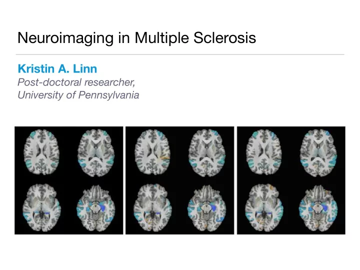
Neuroimaging in Multiple Sclerosis Kristin A. Linn Post-doctoral researcher, University of Pennsylvania Personal Background 2004 - 2008 2009 - 2014 2014 - 2017 Recurse Center Structural MRI 3D array of voxels 2D Grayscale
Neuroimaging in Multiple Sclerosis Kristin A. Linn Post-doctoral researcher, University of Pennsylvania
Personal Background 2004 - 2008 2009 - 2014 2014 - 2017 Recurse Center
Structural MRI 3D array of voxels • 2D Grayscale intensities provide • pixel contrast between different tissue types Standard size approximately • 256 x 256 x 176 voxels 3D voxel Axial slice of a T1 image
Structural MRI in Multiple Sclerosis Used to study lesion accumulation in brain and spinal cord • MS lesion dynamics Inflammation
Structural MRI in Multiple Sclerosis Used to study lesion accumulation in brain and spinal cord • MS lesion dynamics Inflammation Tissue damage and demylenation
Structural MRI in Multiple Sclerosis Used to study lesion accumulation in brain and spinal cord • MS lesion dynamics Inflammation Tissue damage and demylenation Tissue repair and remylenation
Structural MRI in Multiple Sclerosis Used to study lesion accumulation in brain and spinal cord • MS lesion dynamics Inflammation Tissue damage and demylenation Tissue repair and remylenation
Quantifying MS Treatment E ffi cacy • Do chronic lesions show evidence of repair in response to treatment? Pre-treatment Post-treatment
Quantifying MS Treatment E ffi cacy • Do chronic lesions show evidence of repair in response to treatment? Pre-treatment Post-treatment • Initial comparison of average chronic lesion intensity before and after treatment was highly significant
Quantifying MS Treatment E ffi cacy • Do chronic lesions show evidence of repair in response to treatment? Pre-treatment Post-treatment • Statistical challenges : confounding, automatic lesion segmentation, image preprocessing
Preprocessing README 12 steps before analysis • Current analysis is a t-test ! • Each induces errors/uncertainty • that is not easy to account for Typically, researchers ignore • preprocessing uncertainty
Registration Error Image alignment sometimes off by one slice • Pre- treatment: Post- treatment:
Registration Error Pre- treatment: Leads to bias in favor of a treatment effect! Post- treatment:
Registration Error Pre- treatment: Post- treatment:
Registration Error Pre- treatment: Post- treatment:
Moving Forward Many opportunities for statisticians to contribute in meaningful ways • to neuroimaging research Need more sophisticated methods to account for preprocessing errors •
Moving Forward Many opportunities for statisticians to contribute in meaningful ways • to neuroimaging research Need more sophisticated methods to account for preprocessing errors • Steep learning curve (anatomy, physics, engineering) •
Moving Forward Many opportunities for statisticians to contribute in meaningful ways • to neuroimaging research Need more sophisticated methods to account for preprocessing errors • Steep learning curve (anatomy, physics, engineering) • Any methods/models we build must be fast with high-quality, well- • tested software
Acknowledgements Imaging and clinical collaborators: Post-doctoral Advisor: Salim Chahin, M.D. (Penn) Gabrial Pilar, M.D. (Penn) Clyde Markowitz, M.D. (Penn) Matthew Schindler, M.D. (NIH) Emily Acton (Penn) Taki Shinohara, Ph.D.
Happy 75 th Anniversary!
Recommend


More recommend
Explore More Topics
Stay informed with curated content and fresh updates.