
BRIEF COMMUNICATION Nausea, vomiting and diarrhea: An unusual presentation of multiple sclerosis Andrew Szilagyi MD FRCPC , Hyman M Schipper MD FRCPC , Norman Just MD FRCPC A Szilagyi, HM Schipper, N Just. Nausea, vomiting and diar- Nausées, vomissements et diarrhée : tableau rhea: An unusual presentation of multiple sclerosis. Can J Gas- inusité de la sclérose en plaques troenterol 1997;11(4):367-370. The case of a young woman who presented with nausea, vomiting and diarrhea is outlined; the RÉSUMÉ : On présente ici le cas d'une jeune femme présentant des etiology turned out to be a first attack of multiple sclerosis. Plausi- nausées, des vomissements et de la diarrhée. L'étiologie s'est révélée ble mechanisms are discussed. être une première crise de sclérose en plaques. Les mécanismes Key Words: Diarrhea, Multiple sclerosis, Vomiting possiblement en cause sont décrits ici. M CASE PRESENTATION ultiple sclerosis is a multifocal demyelinating disorder occurring with highest incidence in temperate cli- In June 1990 a 33-year old right-handed woman presented to mate zones. It usually affects young adults with subacute on- the emergency room with a two-week history of nausea, set of focal neurological symptoms. The majority (80%) of vomiting and intermittent (one to four times/day) loose wa- patients present with visual, sensory or gait disturbances tery stools without blood. The vomiting and diarrhea were (1,2), whereas older patients (older than 40 years) more not associated with cramps, fever or chills. She experienced commonly exhibit symptoms of progressive myelopathy (2). mild light-headedness, especially on arising, and complained Nausea, vomiting and diarrhea are three of the most com- of mild left neck pain. On the day of admission she vomited mon symptoms encountered in patients seen by both family three times. There were no symptoms of upper respiratory physicians and gastroenterologists. In the vast majority, the tract infection and no history of travel or contact with sub- causes include specific or nonspecific gastrointestinal infec- jects with diarrhea. She is married and had two healthy chil- tions (3). However this symptom complex can be caused by dren by caesarian section. She was last treated with antibiot- neurological disease. The usual neurological etiologies in- ics for sinusitis in 1985. There was no history of alcohol or clude autonomic neuropathies (4,5), but rarely central causes substance abuse. The patient’s mother died of disabling mul- can be seen (6-8). This symptom complex is an unusual pre- tiple sclerosis, and a younger brother was recently diagnosed senting feature and may easily confound the diagnosis of with this demyelinating disease. multiple sclerosis, particularly if focal central nervous system Physical examination at admission disclosed a thin signs are absent. The case of a young woman whose initial woman in no acute distress, with normal vital signs and no symptoms of nausea, vomiting and diarrhea appear to repre- postural changes. The general and neurological examina- sent a first attack of multiple sclerosis is presented. tions were entirely within normal limits. Departments of Neurology and Radiology, and Division of Gastroenterology, Sir Mortimer B Davis Jewish General Hospital, McGill University School of Medicine and Faculty of Medicine, Montreal, Quebec Correspondence and reprints: Dr A Szilagyi, 6000 Côte des Neiges, #100, Montréal, Québec H3S 1Z8. Telephone 514-739-7370, fax 514-340-8282 Received for publication May 31, 1996. Accepted November 14, 1996 Can J Gastroenterol Vol 11 No 4 May/June 1997 367
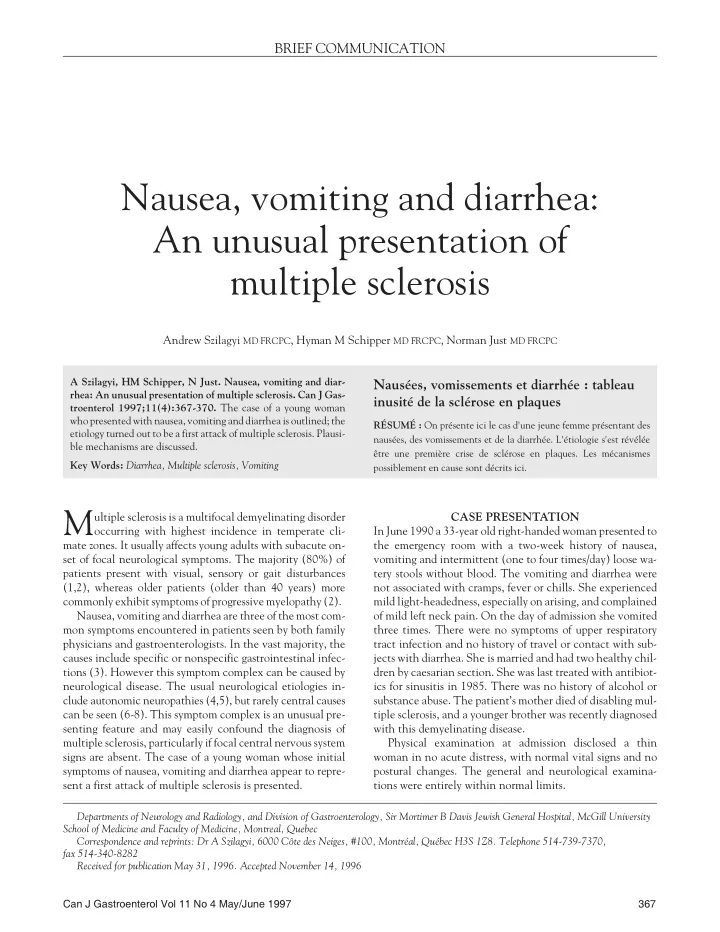
Szilagyi et al Figure 1) Left Magnetic resonance imaging (MRI) shows axial T 2 weighted images of L occipital hyperintense subcortical lesion (arrow). Above Sagittal proton density image through the corpus callosum shows a small lesion at the callososeptal interface (arrow). MRI findings strongly support the diagnosis of multiple sclerosis Hematocrit was 36.3%, white blood cell count 5.3 x 10 9 /L, as a lesion in the corpus callosum (Figure 1). A lumbar punc- mean cell volume 85.6 fL, blood glucose 4.5 mmol/L, blood ture revealed normal cells, protein and glucose, and negative urea nitrogen 2.9 mmol/L and creatinine 84 � mol/L. Elec- bacterial microbiology. However, oligoclonal bands were trolytes and liver enzymes were normal. A free thyroxine in- present, supporting the diagnosis of multiple sclerosis. Soma- dex and thyroid stimulating hormone were normal. tosensory evoked potentials showed interference with con- Urinalysis and urine cultures were normal. A Venereal Dis- duction in the left cerebral hemisphere consistent with ease Research Laboratory test for syphilis was negative. An lesions in the left subcortical white matter. electrocardiogram test was normal. The patient improved somewhat on dimenhydrinate sup- The patient was admitted for further evaluation. A gas- positories and was discharged after eight days of hospitaliza- troenterological work-up consisting of stool occult blood, tion. However, she was readmitted with worsening nausea cultures, sensitivity, ova and parasites were all negative. and vomiting six days later; she also complained of diffuse Gastroscopy and an upper gastrointestinal small bowel nonthrobbing headaches. She was started on intravenous follow-through study were also normal. A colonoscopy to methylprednisolone sodium 250 mg q6h and intravenous splenic flexure with biopsy of normal-looking mucosa dis- promethazine hydrochloride 25 mg q4h. During this second closed mild nonspecific edema and occasional inflammatory hospitalization, an episode of right hand and right thigh cells in the rectosigmoid. A liquid gastric emptying scan paresthesia lasted two to three days. A repeat CT scan of the showed 73% retention after 30 mins (normal less than 50%). head showed a prominent demyelinating plaque in the left A solid gastric emptying scan was normal. corticomedullary region. She gradually improved and was Because symptoms persisted and nausea was a prominent discharged on oral promethazine hydrochloride 26 days after feature, and because of the family history, neurological con- the second hospitalization. Promethazine hydrochloride was sultation was obtained. Clinical evaluation and a Bàràny test discontinued three months later, without a relapse of gastro- were normal. A noncontrast computed tomographic (CT) intestinal symptoms. She has been well without gastrointes- scan of the head revealed a small hypodense lesion in the left tinal or neurological symptoms for the past five years. subcortical white matter. A follow-up double dose delayed DISCUSSION contrast CT scan of the head revealed two diffusely enhanc- ing left parietal white matter lesions compatible with multi- Gastrointestinal manifestations of multiple sclerosis include ple sclerosis. Magnetic resonance imaging (MRI) with transfer and transport dysphagia (9), nausea, vomiting (2), T 2 -weighted images disclosed a much wider involvement of Brun's syndrome (vertigo, vomiting, headache and visual the subcortical white matter, with lesions in the left parietal, disturbances with positional change of the head) (2,10), left occipital, right parietal and right optic radiation, as well constipation and fecal incontinence (11,12). Inflammatory 368 Can J Gastroenterol Vol 11 No 4 May/June 1997
Recommend
More recommend