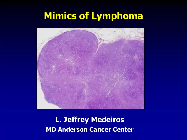

Mimics of Lymphoma L. Jeffrey Medeiros MD Anderson Cancer Center
Mimics of Lymphoma Outline Progressive transformation of GCs Infectious mononucleosis Kikuchi-Fujimoto disease Castleman disease Metastatic seminoma Metastatic nasopharyngeal carcinoma Thymoma Myeloid sarcoma
Progressive Transformation of Germinal Centers (GC) Clinical Features Occurs in 3-5% of lymph nodes Any age: 15-30 years old most common Usually localized Cervical LNs # 1 Uncommonly patients can present with generalized lymphadenopathy involved by PTGC Fever and other signs suggest viral etiology
Progressive Transformation of GCs Different Stages Early Mid-stage
Progressive Transformation of GCs Later Stage
Progressive Transformation of GCs IHC Findings CD20 CD21 CD10 BCL2
Progressive Transformation of GCs Histologic Features Often involves small area of LN Large nodules (3-5 times normal) Early stage: Irregular shape Blurring between GC and MZ Later stages: GCs break apart Usually associated with follicular hyperplasia Architecture is not replaced
Differential Diagnosis of PTGC NLPHL Nodules replace architecture LP (L&H) cells are present Lymphocyte- Nodules replace architecture rich classical Small residual germinal centers HL, nodular RS+H cells (CD15+ CD30+ LCA-) variant Follicular Numerous follicles lymphoma Back-to-back Into perinodal adipose tissue Uniform population of neoplastic cells PTGC – differential dx
Nodular Lymphocyte Predominant HL CD20 CD3 NLPHL
Lymphocyte-rich Classical HL Nodular variant CD20 CD15 LRCHL
Progressive Transformation of GCs BCL2+ is not evidence of follicular lymphoma BCL2 BCL6
Practical Questions Is PTGC associated with future risk of NLPHL? Statistically no but … Do I comment on possible relationship to NLPHL in the pathology report? Never Is PTGC one lesion? Probably not - may be more than 1 type
Infectious Mononucleosis Basic Facts Caused by Epstein-Barr virus (HHV-4) Spread by contact with human secretions Age of contact depends on living conditions Poor - < 3 years Good - 10-19 years Incubation period is 2-5 weeks First week Humoral antibody response Second week Cellular immune response
Infectious Mononucleosis Clinical Features Fever, pharyngitis, lymphadenopathy (50%) Lymphocytosis with atypical lymphocytes Less common: Hepatosplenomegaly Tonsillitis Thrombocytopenia Anemia Skin rash Rarely IM can occur in the elderly
Infectious Mononucleosis Tonsillitis
Infectious Mononucleosis Lymphocytosis Hal Downey, PhD (1877-1959)
Infectious Mononucleosis
Infectious Mononucleosis RS-like cell
Infectious Mononucleosis CD20 CD3 EBER CD30
Infectious Mononucleosis Histologic Features Marked expansion of the paracortex Often some preservation of architecture Spectrum of cells Many immunoblasts RS-like cells +/- Necrosis common Many EBER+ cells Follicular hyperplasia is common
Differential Diagnosis of Infectious Mononucleosis CMV Can resemble IM histologically lymphadenitis CMV inclusions +/- EBV absent Large B-cell Architecture replaced lymphoma Monotonous cell population EBV negative (usually) Monoclonal Anaplastic large Sinusoidal (common) cell lymphoma Hallmark cells, ALK+ Monoclonal Classical No spectrum of cell types Hodgkin RS+H cells: CD15+ LCA- lymphoma
Kikuchi-Fujimoto Lymphadenitis Clinical Features First described in 1972 in Japan A.K.A. histiocytic necrotizing lymphadenitis Median age 30 years (wide range) Female predominance Cervical LNs # 1 Patients present with: Moderate fever, chills Myalgias +/- Masahiro Kikuchi, MD
Kikuchi-Fujimoto Lymphadenitis Paracortical and wedge-shaped infiltrate
Kikuchi-Fujimoto Lymphadenitis Proliferative Phase
Kikuchi-Fujimoto Lymphadenitis Necrotizing Necrotizing and Xanthomatous
Kikuchi-Fujimoto Lymphadenitis Necrotic and proliferative stages CD123
Kikuchi-Fujimoto Lymphadenitis Histologic Features Overall architecture preserved Paracortical; Patchy necrosis + / - Increased histiocytes; often C-shaped Increased plasmacytoid dendritic cells (CD123+) No granulocytes; no (or rare) plasma cells Follicular hyperplasia +/- 3 phases: Necrotizing Proliferative Xanthomatous
Kikuchi-Fujimoto Lymphadenitis Immunophenotype Numerous histiocytes CD68+, CD123+, lysozyme+, MPO+ Many T-cells CD8 > CD4 CD30+ immunoblasts Ki-67 can be high Ki67
Kikuchi-Fujimoto Lymphadenitis IHC Findings CD3 CD68 MPO CD20
Differential Diagnosis of Kikuchi-Fujimoto Lymphadenitis SLE Can be identical to K-F lymphadenitis Hematoxylin bodies +/- Infectious Different quality of necrosis lymphadenitis (coagulative with polys) Infarcted Ghosts of tumor cells lymphoma Immunostains highlight dead cells Large B-cell Only proliferative phase of K-F lymphoma Immunophenotype helps
Castleman Disease Clinical Pathological Unicentric Hyaline-vascular variant Plasma cell variant (HHV-8-) Multicentric Plasma cell variant (HHV-8+)
Hyaline-vascular Castleman Disease Clinical Features 90% of all cases of unicentric CD Almost any age (8-70 yrs) Usually asymptomatic Small or very large mass (up to 16 cm) Usually above the diaphragm Mediastinum is # 1 site Surgical excision is optimal therapy
Hyaline-vascular Castleman Disease Histologic Features Follicular Large follicles “Twinning” “Onion - skin” mantle zones Lymphocyte depletion of germinal centers Hyaline-vascular lesions Interfollicular This can be predominant (stroma-rich) Numerous high endothelial venules Actin+, CD68+, CD21+
Hyaline-vascular Castleman Disease
Hyaline-vascular Castleman Disease Twinning Hyaline-vascular lesion CD – hyaline-vascular variant
Hyaline-vascular Castleman Disease Stroma Rich CD21 CD – hyaline-vascular variant
32 cases analyzed by HUMARA assay 25 / 32 cases were monoclonal 22 / 29 hyaline vascular variant 3 / 3 plasma cell variant 3 cases had clonal karyotypes No IGH or TCRG or TCRB rearrangements Hyaline vascular CD may be a neoplasm of stromal cells Mod Pathol 27: 823, 2014
Differential Diagnosis of Hyaline-vascular Castleman Disease Follicular hyperplasia No hyaline-vascular lesions No lymphocyte depletion No interfollicular vascularity Follicular lymphoma Follicles are numerous and monotonous No lymphocyte depletion No interfollicular vascularity Mantle cell CD5+ cyclin D1+ lymphoma, mantle zone pattern Plasma cell variant Marked plasmacytosis CD Can have H-V follicles
Plasma Cell CD (Unicentric) Clinical Features 10% of unicentric CD Almost any age One or multiple small lymph nodes Systemic symptoms in a subset (? multicentric CD)
Plasma Cell CD (Unicentric) Histologic and Immunophenotypic Features Interfollicular sheets of plasma cells Sinuses usually patent Follicles have some H-V lesions +/- Polytypic plasma cells and B-cells Human herpes virus 8 (KSHV) -
Plasma Cell CD (Unicentric)
Differential Diagnosis of Plasma Cell CD (Unicentric) Rheumatoid arthritis Grossly smaller No H-V lesions Plasmacytoma Replacement of LN architecture Multicentric CD HHV-8+ (usually HIV+)
Multicentric Castleman Disease Clinical Features Usually associated with systemic symptoms Often associated with HIV infection Lymphadenopathy – 100% of patients Hepatosplenomegaly, effusions, skin rash +/- Laboratory Elevated ESR, anemia, thrombocytopenia Polyclonal hypergammaglobulinemia
Multicentric Castleman Disease Histologic and Immunophenotypic Features Similar to unicentric plasma cell variant Interfollicular sheets of plasma cells Atypical plasma cells Follicles show H-V lesions Blurring of boundary between germinal centers and mantle zones HHV-8+, EBV+/- Plasma cells can be monotypic
Multicentric Castleman Disease HIV Positive HHV-8
Multicentric Castleman Disease Presence of “Microlymphoma” kappa HHV-8 lambda
Differential Diagnosis of Multicentric Castleman Disease Unicentric Unicentric plasma cell HHV-8- variant No HIV infection Hyaline-vascular HV lesions variant Big follicles Interfollicular vascularity Peripheral T-cell Architecture effaced lymphoma Monoclonal T-cell population
POEMS Syndrome Polyneuropathy, organomegaly, endocrinopathy, M protein, skin changes Paraneoplastic syndrome caused by elevated angiogenic and inflammatory cytokines Associated with underlying plasma cell dyscrasia 95% lambda Often osteosclerotic 50% of patients have Castleman disease, plasma cell variant
Multicentric Castleman Disease POEMS Syndrome
TAFRO Syndrome Thrombocytopenia, Anasarca, Fever, Reticulin fibrosis in BM, and Organomegaly Also known as Castleman-Kojima disease Most common in Japan Women most often affected ? Cytokine storm attributable to IL-6 and VEGF J Clin Exp Hematop 53: 95, 2103
Seminoma Clinical Features Most common germ cell tumor of testis Age range: 30-45 years 80-90% have a palpable mass Often no symptoms; testicular pain ~20% Laboratory tests: LDH HCG (~10%) AFP negative 75% of pts have stage I (localized) disease Metastases to: retroperitoneal LNs, lungs
Metastatic Seminoma to LN PLAP
Recommend
More recommend