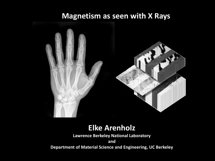

Magnetism as seen with X Rays Elke Arenholz Lawrence Berkeley National Laboratory and Department of Material Science and Engineering, UC Berkeley 1
What to expect: + Magnetic Materials Today + Magnetic Materials Characterization Wish List + Soft X-ray Absorption Spectroscopy – Basic concept and examples + X-ray magnetic circular dichroism (XMCD) - Basic concepts - Applications and examples - Dynamics: X-Ray Ferromagnetic Resonance (XFMR) + X-Ray Linear Dichroism and X-ray Magnetic Linear Dichroism (XLD and XMLD) + Magnetic Imaging using soft X-rays + Ultrafast dynamics 2
Magnetic Materials Today Magnetic materials for energy applications Magnetic nanoparticles for biomedical and environmental applications Magnetic thin films for information storage and processing 3
Permanent and Hard Magnetic Materials Controlling grain and domain structure on the micro- and nanoscale Engineering magnetic anisotropy on the atomic scale 4
Magnetic Nanoparticles Optimizing magnetic nanoparticles for biomedical Tailoring magnetic applications nanoparticles for environmental applications 5
Magnetic Thin Films Magnetic domain structure on the nanometer scale Magnetic coupling at interfaces Unique Ultrafast magnetic magnetization phases at reversal interfaces dynamics GMR Read Head Sensor 6
Magnetic Materials Characterization Wish List + Sensitivity to ferromagnetic and antiferromagnetic order + Element specificity = distinguishing Fe, Co, Ni, … + Sensitivity to oxidation state = distinguishing Fe 2+ , Fe 3+ , … + Sensitivity to site symmetry, e.g. tetrahedral, T d; octahedral, O h + Nanometer spatial resolution + Ultra-fast time resolution Soft X-Ray Spectroscopy and Microscopy 7
Spectroscopy Light/Photon Source Monochromator Sample sample intensity intensity wavelength, photon energy wavelength, photon energy 8
Soft X-Ray Spectroscopy ( h 500-1000eV, 1-2nm) transmitted intensity I t / I 0 1.0 0.9 0.8 Electron Storage Ring Monochromator 0.7 EY_TM.opj Sample 0.6 770 780 790 800 photon energy (eV) 9
X-Ray Absorption Detection Modes transmitted intensity I t / I 0 1.0 0.9 Photons 0.8 absorbed 0.7 EY_TM.opj 0.6 770 780 790 800 photon energy (eV) mean 6 free electron yield I e / I 0 path 5nm 4 Electrons generated 2 EY_TM.opj 0 770 780 790 800 photon energy (eV) J. Stöhr, H.C. Siegmann, Magnetism (Springer) Electron yield: + Absorbed photons create core holes subsequently filled by Auger electron emission + Auger electrons create low-energy secondary electron cascade through inelastic scattering + Emitted electrons probability of Auger electron creation absorption probability 10
Soft X-Ray Absorption – Probing Depth Element 10eV below L 3 at L 3 40 eV above L 3 1/ [nm] 1/ [nm] 1/ [nm] Fe 550 17 85 Co 550 17 85 Ni 625 24 85 // // // 3nm Mn Fe Co Ni Ru 1.0 15nm NiFe 3nm I t / I 0 Ru luminescence_BL402_Apr2012.opj 15nm CoFe 0.8 15nm MnIr // // // 5nm 860 880 640 660 700 720 780 800 Ru photon energy (eV) 10-20 nm layer thick films supported by substrates transparent to soft X rays 11
X-Ray Absorption Detection Modes and Probing Depth e Mn Fe Co Ni 1.0 1.0 1.0 1.0 3nm Ru I t / I 0 15nm 0.8 0.8 NiFe 0.8 0.8 3nm Ru electron yield (arb. units) 1.1 15nm CoFe 1.0 1.0 1.4 luminescence_BL402_Apr2012.opj 15nm MnIr 1.0 1.2 5nm Ru 0.9 1.0 0.9 640 650 660 710 720 780 790 850 860 870 photon energy (eV) photon energy (eV) photon energy (eV) photon energy (eV) + Electron sample depth: 2-5 nm in Fe, Co, Ni 60% of the electron yield originates form the topmost 2-5 nm 12
X-Ray Absorption Fundamentals Experimental Concept: Monitor reduction in X-ray flux transmitted through sample as function of photon energy E Charge state of absorber Fe 2+ , Fe 3+ Valence Symmetry of lattice site of absorber: O h , T d states Sensitive to magnetic order Absorption probability: X-ray energy, X-ray polarization, experimental geometry 2 p 3/2 L 3 Core level Atomic species of absorber Fe, Co, Ni, …. 2 p 1/2 L 2 13
X-Ray Absorption Fundamentals Experimental Concept: Monitor reduction in X-ray flux transmitted through sample as function of photon energy E Charge state of absorber Fe 2+ , Fe 3+ Valence Symmetry of lattice site of absorber: O h , T d states Sensitive to magnetic order Absorption probability: X-ray energy, X-ray polarization, experimental geometry 2 p 3/2 L 3 Core level Atomic species of absorber Fe, Co, Ni, …. 2 p 1/2 L 2 14
‘White Line’ Intensity Fe valence states 2 p 3/2 L 3 core level 2 p 1/2 L 2 Intensity of L 3,2 resonances is proportional to number of d states above the Fermi level, i.e. number of holes in the d band. 15
‘White Line’ Intensity Fe valence states 2 p 3/2 L 3 core level 2 p 1/2 L 2 Intensity of L 3,2 resonances is proportional to number of d states above the Fermi level, i.e. number of holes in the d band. 16
‘White Line’ Intensity Co valence states 2 p 3/2 L 3 core level 2 p 1/2 L 2 Intensity of L 3,2 resonances is proportional to number of d states above the Fermi level, i.e. number of holes in the d band. 17
‘White Line’ Intensity Ni valence states core level 2 p 3/2 L 3 2 p 1/2 L 2 Intensity of L 3,2 resonances is proportional to number of d states above the Fermi level, i.e. number of holes in the d band. 18
‘White Line’ Intensity Cu valence states core level 2 p 3/2 L 3 2 p 1/2 L 2 Intensity of L 3,2 resonances is proportional to number of d states above the Fermi level, i.e. number of holes in the d band. 19
X-Ray Absorption Fundamentals Experimental Concept: Monitor reduction in X-ray flux transmitted through sample as function of photon energy E Charge state of absorber Fe 2+ , Fe 3+ , … Valence Symmetry of lattice site of absorber: O h , T d states Sensitive to magnetic order Absorption probability: X-ray energy, X-ray polarization, experimental geometry 2 p 3/2 L 3 Core level Atomic species of absorber Fe, Co, Ni, …. 2 p 1/2 L 2 20
X-ray Absorption – Valence State Influence of the charge state of the absorber N. Telling et al ., Appl. Phys. Lett. 95, 163701 (2009) J.-S. Kang et al., Phys. Rev. B 77, 035121 (2008) 21
X-Ray Absorption – Configuration Model Ni 2+ in NiO: 2 p 6 3 d 8 → 2 p 5 3 d 9 Configuration model, e.g. L edge absorption : + Excited from ground/initial state configuration, 2 p 6 3 d 8 to exited/final state configuration, 2 p 5 3 d 9 + Omission of all full subshells (spherical symmetric) + Takes into account correlation effects in the ground state as well as in the excited state + Leads to multiplet effects/structure http://www.anorg.chem.uu.nl/CTM4XAS / 2 S +1 L J. Stöhr, H.C. Siegmann, Magnetism (Springer) 22
X-Ray Absorption Fundamentals Experimental Concept: Monitor reduction in X-ray flux transmitted through sample as function of photon energy E Charge state of absorber Fe 2+ , Fe 3+ Valence Symmetry of lattice site of absorber: O h , T d states Sensitive to magnetic order Absorption probability: X-ray energy, X-ray polarization, experimental geometry 2 p 3/2 L 3 Core level Atomic species of absorber Fe, Co, Ni, …. 2 p 1/2 L 2 23
Sensitivity To Site Symmetry: Ti 4+ L 3,2 3 d orbitals PbZr 0.2 Ti 0.8 O 2 e e t 2 t 2 XA (arb. units) J. Stöhr, H.C. Siegmann, PZT_XA.opj + Electric dipole transitions: d 0 2 p 5 3 d 1 Magnetism (Springer) 455 460 465 470 + Crystal field splitting 10 Dq acting on 3 d orbitals: photon energy (eV) Tetragonal symmetry: Octahedral symmetry: e orbitals towards ligands higher energy e orbitals b 2 = d xy , e = d yz , d yz t 2 orbitals b 1 = d x2 y2 , a 1 = d 3z2 r2 t 2 orbitals between ligands lower energy 24
X-Ray Absorption Lattice Symmetry TiO 2 rutile Influence of lattice site symmetry at the absorber x- ray absorption O h anatase TiO 2 D 2h G. Van der Laan Phys. Rev. B 41, 12366 (1990) 25
X-Ray Absorption Fundamentals Experimental Concept: Monitor reduction in X-ray flux transmitted through sample as function of photon energy E Charge state of absorber Fe 2+ , Fe 3+ Valence Symmetry of lattice site of absorber: O h , T d states Sensitive to magnetic order Absorption probability: X-ray energy, X-ray polarization, experimental geometry 2 p 3/2 L 3 Core level Atomic species of absorber Fe, Co, Ni, …. 2 p 1/2 L 2 26
X-Ray Absorption Fundamentals Experimental Concept: Monitor reduction in X-ray flux transmitted through sample as function of photon energy E Charge state of absorber Fe 2+ , Fe 3+ Valence Symmetry of lattice site of absorber: O h , T d states Sensitive to magnetic order Absorption probability: X-ray energy, X-ray polarization, experimental geometry 2 p 3/2 L 3 Core level Atomic species of absorber Fe, Co, Ni, …. 2 p 1/2 L 2 27
Recommend
More recommend