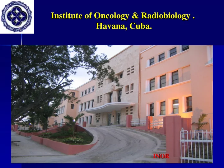

Institute of Oncology & Radiobiology . Institute of Oncology & Radiobiology . Havana, Cuba. Havana, Cuba. INOR INOR 1 1
“Transition from 2 Transition from 2- -D to 3 D to 3- -D conformal radiotherapy D conformal radiotherapy “ in high grade gliomas gliomas: our experience in Cuba : our experience in Cuba” ” in high grade Chon. I, MD - - Chi. D, MD - - Alert.J, MD- - Alfonso. R, PhD.- - Ropero. . R, MD. Chon. I, MD Chi. D, MD Alert.J, MD Alfonso. R, PhD. Ropero R, MD. Department of Radiotherapy Department of Radiotherapy Institute of Oncology & Radiobiology . Havana, Cuba. Institute of Oncology & Radiobiology . Havana, Cuba.
� The aims of 3D The aims of 3D- -CRT are to achieve conformity of CRT are to achieve conformity of � the high dose region to the target volume and the high dose region to the target volume and consequently to reduce the dose reaching the consequently to reduce the dose reaching the surrounding normal tissues. This should reduce both surrounding normal tissues. This should reduce both acute and late morbidity. If the adverse effects of acute and late morbidity. If the adverse effects of treatment can be reduced in this way, the dose of the treatment can be reduced in this way, the dose of the target volume can be increased with the expectation target volume can be increased with the expectation of improving survival. of improving survival. � It is now the standard practice in developed It is now the standard practice in developed � countries, treating many types of tumours tumours with with countries, treating many types of curative intent. curative intent. 3 3
THE GOALS OF THE PRESENT STUDY ARE: THE GOALS OF THE PRESENT STUDY ARE: Firstly, to compare the effects of radiation dose- - Firstly, to compare the effects of radiation dose escalation in adult patients, treated with third escalation in adult patients, treated with third dimension conformal radiation therapy (3- -D CRT) D CRT) dimension conformal radiation therapy (3 with those patients who had just the second dimension with those patients who had just the second dimension radiation therapy (2- -D RT). All patients have high D RT). All patients have high radiation therapy (2 grade gliomas gliomas. . grade Secondly, to show the benefits of third dimension Secondly, to show the benefits of third dimension conformal radiation therapy (3- -D CRT) as the D CRT) as the conformal radiation therapy (3 treatment of choice for malignant gliomas gliomas in the treatment of choice for malignant in the postoperative stage. postoperative stage. 4 4
Patients and and Methods Methods: : Patients � A total A total of of 45 45 patients patients with with supratentorial supratentorial high grade grade high � gliomas were were included included from from 2004 2004 to to 2007 . 2007 . The The treatments treatments gliomas were performed performed in in our our radiotherapy radiotherapy department department . . were � The The inclusion inclusion/ /exclusion exclusion criteria criteria were were: : � -Anaplastic Anaplastic Astrocytoma (AA) and Glioblastoma - Astrocytoma (AA) and Glioblastoma Multiforme (GBM) histology histology. . Multiforme (GBM) -Karnofsky Karnofsky Performance Score (KPS) ≥ ≥ 70. 70. - Performance Score (KPS) -18 18- -65 65 years years old. . - old -Total Total or or subtotal macroscopic macroscopic surgical resection. . - subtotal surgical resection -No No previous previous chemotherapy/ / inmunotherapy inmunotherapy treatment. . - chemotherapy treatment -Informed Informed consent obtained. . - consent obtained 5 5
Control Group Group Control � DTT : 60 DTT : 60 Gy Gy � (2 Gy Gy x 5d / wk wk during 6 weeks weeks) ) (2 x 5d / during 6 � The The total total treated treated volume volume was was: tumor + : tumor + oedema oedema +3 +3- -4cm 4cm � of margins margins of local fields fields) ) ( local 2D Conventional Conventional Radiotherapy Radiotherapy ( 2D � � 7 7
Prospective Group Group Prospective DTT : 66 - - 70 Gy Gy (1,8 Gy Gy x 5d/wk wk during 7- -8 8 weeks weeks) ) • DTT : 66 70 (1,8 x 5d/ during 7 • • Treatment Treatment Volumes (ICRU 50 & 62): Volumes (ICRU 50 & 62): • GTV: enhanced enhanced contrast lesion defined by CT or or MRI. * GTV: contrast lesion defined by CT MRI. * *CTV 1: enhanced enhanced contrast lesion + the perilesional edema + + 3- -4 4 cm cm of of *CTV 1: contrast lesion + the perilesional edema 3 margins. . margins *CTV 2: enhanced enhanced contrast lesion + 2cm 2cm of of margins. . *CTV 2: contrast lesion + margins *PTV 1: CTV1+ 10 + 10- -15mm margins when technique is uncertain. 15mm margins when technique is uncertain. *PTV 1: CTV1 *PTV2: CTV2 CTV2 + margin of 10 + margin of 10- -15mm when technique is uncertain. 15mm when technique is uncertain. *PTV2: Level 2 (the the European Dynarad Consortium) ) of of • Level 2 ( European Dynarad Consortium 3D Conformal Conformal 3D • Radiotherapy Radiotherapy 8 8
2D CONVENTIONAL RADIOTHERAPY. . Conventional Simulator Conventional Simulator � � (Beam ( Beam geometry geometry determined determined by by fluoroscopic fluoroscopic simulation simulation) ) Immobilization: Velcro strap, head support Immobilization: Velcro strap, head support � � 2D treatment planning systems: 2D treatment planning systems: � � – Theraplan Plus (Basic, non image based) – Theraplan Plus (Basic, non image based) Treatment Machine : Treatment Machine : � � – – Co Co 60 Theratronics 60 Theratronics Phoenix Phoenix 9 9
3D Conformal Conformal Radiotherapy Radiotherapy 3D Imaging Equipment Imaging Equipment � � (multi- -slice CT slice CT- - Scanner) (multi Scanner) Immobilization: thermoplastic mask Immobilization: thermoplastic mask � � 3D image based treatment planning systems: 3D image based treatment planning systems: � � – Theraplan Plus (Advanced) – Theraplan Plus (Advanced) – – PrecisePLAN PrecisePLAN V. 2.12 V. 2.12 Treatment Machine Treatment Machine � � – – 2 Elekta 2 Elekta Precise linacs Precise linacs (MLC & EPID) & EPID) (MLC � � Network R&V System R&V System � � 10 10 and Networking and Networking
3- -D D 3 2- -D D 2 Treatment portals portals were were determined determined Treatment � � Treatment planning is is based Treatment planning based on � � on , where where based on bony bony landmarks landmarks, based on designing beam designing beam 3- -D D anatomy, , 3 anatomy the target was the tumor and and the target was the tumor geometries and treatment portals geometries and treatment portals peritumoral tissue peritumoral tissue. . Critical Critical structures structures according to according to the the extension extension of of target target were avoided avoided or or not not. . were and risk risk structures structures. . and Limited information information was was obtained obtained Limited � � The plan evaluation was done The plan evaluation was done � � about about isodose isodose distributions distributions such such as as through through the the 2D 2D isodose isodose curves curves for for the minimum and the maximum the minimum and the maximum Multiple Plannar Reconstruction Multiple Plannar Reconstruction tumor and tumor and normal tissues normal tissues doses doses (MPR), 3D isosurface (MPR), 3D isosurface and and Dose Dose received received Volumen Histogram Histogram (DVH). (DVH). Volumen Evaluation plan plan consisted consisted only only in in the the Evaluation � � 3- -D D treatment treatment is is verified verified comparing comparing 3 � � examination of examination of one one or or a a very very few few DRR ( DRR (from from the the 3D CT data), 3D CT data), with with cross- -sectional sectional images images. . cross the portal portal images images acquired acquired by by films films the 2- -D D treatment was verified 2 treatment was verified or or EPIDs EPIDs. . � � comparing port port films films with with simulator simulator comparing films. . films 11 11
CLASSIFICATION OF CONFORMAL THERAPY ACCORDING TO THE METHODOLOGY AND TOOLS ASSOCIATED WITH EACH STEP OF THE PROCEDURE ( IAEA TECDOC 1588 )
Recommend
More recommend