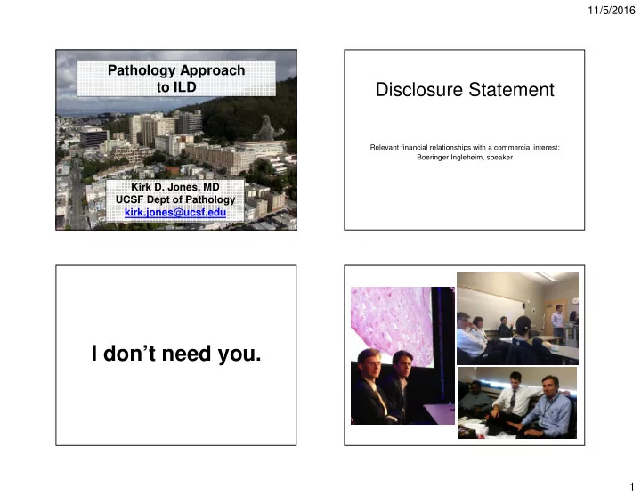

11/5/2016 Pathology Approach to ILD Disclosure Statement Relevant financial relationships with a commercial interest: Boeringer Ingleheim, speaker Kirk D. Jones, MD UCSF Dept of Pathology kirk.jones@ucsf.edu I don’t need you. 1
11/5/2016 Normal Lung Overview • The lung is divided into numerous lobular units that have a characteristic appearance. • Normal lung • Arteries run with airways. • Patterns of fibrosis • Veins present in interlobular septa. • A little on granulomas • Lymphatics in bronchovascular bundles, interlobular septa, and pleura. 2
11/5/2016 3
11/5/2016 Pattern of Fibrosis Thinking about Fibrosis The distribution of the fibrosis will often correlate with the • nature of the injury. • Usually in patients with chronic or insidious Bronchiolocentric fibrosis – tends to occur in diseases • with inhalation injury (HP, RB, fume) or bronchiolar disease. inflammation (CTD) • Often observed on CT scan as reticulation NSIP – tends to occur in diseases with diffuse alveolar • inflammation (autoimmune CTD, drug reaction, HP) (possibly with traction bronchiectasis or honeycombing) or nodules (when UIP – odd peripheral distribution pattern. Possibly related • bronchiolocentric) to aberrent sensecence with most distal cells either more predisposed to stretch injury, or least likely to be replenished. 4
11/5/2016 5
11/5/2016 Bronchiolocentric Fibrosis • Look for lace-like central regions (fireworks) of peribronchiolar metaplasia • Think about inhaled diseases (HP, RB, fume inhalation injury) and diseases with small airway inflammation (aCTD) Nonspecific Interstitial Pneumonia (NSIP) • Diffuse alveolar septal thickening by either inflammation (cellular NSIP) or fibrosis (NSIP- fibrosis) • Can be variable, but should show fibrous thickening of the alveolar septa in peribronchiolar, subpleural, and midzones of the lobule. 6
11/5/2016 Dusty Cobweb Fibrosis NSIP Pattern • Look for variable but diffuse alveolar septal thickening (dusty cobweb) by fibrosis or inflammation • Look for additional clues to help decide the differential (lymphoid aggregates, granulomas, pleuritis, vessel thickening). Term by Kevin Leslie If my pathologist tells me the biopsy shows NSIP, then my job has only just begun. 7
11/5/2016 8
11/5/2016 Clues for DAD UIP Pattern • Fibrosis beginning at the periphery of the lobule • A compatible clinical history • Temporal and spatial heterogeneity • Diffuse alveolar septal thickening or effacement • Temporal (“HORN”) by collapse, edema, and alveolar filling Honeycombing, old (dense collagen) fibrosis, recent (fibroblast foci) • • Hyaline membranes fibrosis, and normal • Distal airway squamous metaplasia • Spatial – worse subpleural, paraseptal, and basilar 9
11/5/2016 10
11/5/2016 11
11/5/2016 12
11/5/2016 The “Tip Test” • Since UIP shows peripheral lobular accentuation of fibrosis, the very tip of the surgical biopsy is often obliterated by fibrosis (often with overlying fatty metaplasia of the pleura) • NSIP tends to show normal alveolar architecture (with thickened septa) at the tip of the biopsy. 13
11/5/2016 OP or FF? UIP Pattern • Fibroblastic Foci • Organizing Pneumonia – Crescentic (frequently) – Rounded (usually) – Collagen on one side – Air on most sides – Location - interstitium – Location - airspace – Sessile – Polypoid – Not branched • When there is pure temporal heterogeneity, the – Branching diagnosis is almost certain • Use the “Tip Test” • Rare cases of HP, CTD may show UIP pattern Look for central scarring and granulomas in HP • Look for increased lymphoid aggregates and lack of normal (NSIP instead) in CTD • Organizing Pneumonia Organizing Pneumonia • Clinically acute or subacute presentation. • The appearance of this disorder looks like something got messed up (a pneumonia of • Patchy opacifications on CT. some sort) and is now being cleaned up (organizing) • The body’s method of cleaning up is via • Pathology findings will mirror CT findings with granulation tissue (fibroblasts and small filling of alveolar spaces. vessels with some inflammatory cells). 14
11/5/2016 BOOP Organizing Pneumonia Alveolar duct Alveoli Bronchiole Granulation tissue polyp Bronchiolitis obliterans 15
11/5/2016 Organizing Pneumonia 16
11/5/2016 Acute Fibrinous Organizing Pneumonia • Polypoid plugs of fibrin (pink slightly granular to fibrillar material) often with flattened cells at the periphery • Can look very similar to eosinophilic pneumonia, so make sure you look for eosinophils when you see AFOP Clues for OP • Correlates with CT and clinical • Rounded branching plugs (Nordy) • Blue with spindly cells (OP) • Pink with few cells (AFOP) 17
11/5/2016 Granulomas Granuloma – Soft Findings • Tightly packed cells, rounded, coalescing, present along lymphatic routes, lymphocytes exclude the interior = sarcoidosis • Finding granulomas on a biopsy can help make • Tightly packed, rounded, singletons, random or several diagnoses, so it is important to bronchiolocentric, lymphocytes in interior = MAC, recognize what they look like hot-tub lung • Rounded aggregates of histiocytes (tissue • Loose, formed of only a few macrophages and giant cells, may show intracytoplasmic cholesterol clefts, macrophages) often with multinucleate giant bronchiolocentric often = HP cells • Associated with neutrophils, big floppy giant cells = aspiration 18
11/5/2016 Sarcoidosis? 19
11/5/2016 Hot-tub lung (M. avium) Hypersensitivity Pneumonia 20
11/5/2016 Hypersensitivity Pneumonia • Cases we have observed: Feathers: Pets, Farm animal, Duvet, Pillow, Jacket. • Molds: Work freezer, Man-Cave, Sleep number • mattress, Hay, Orchid bark Mycobacteria: Indoor spa, shower • Machine oil • ? Central valley: Almond dust? • Courtesy of Rick Webb, MD 21
11/5/2016 Venous injection of crushed tablets Aspiration Illustrative Case • 62-year-old man with severe pulmonary fibrosis • Prior biopsy with UIP pattern • Now undergoing bilateral lung transplant 22
11/5/2016 Pathologic Pattern • Usual interstitial fibrosis • Marked fibrosis with honeycombing • Patchy involvement of lung • Fibroblast foci present • ?Features suggesting alternate diagnosis? 23
11/5/2016 Pathologic Diagnosis • Interstitial fibrosis, UIP pattern, with bronchiolocentric fibrosis and chronic inflammation, and poorly formed granulomas. • Most consistent with chronic hypersensitivity pneumonia. Final Diagnosis • Familial Interstitial Fibrosis • Telomerase mutation (TERT gene) • With superimposed hypersensitivity pneumonia 24
11/5/2016 Take Away Message I still think that it is good that we are each in our • respective career – and that the best way to make a diagnosis is through multidisciplinary discussion. However, the longer I do this, the more value I find in • knowing at least some of what the other team members do. It is my hope that I have effectively communicated a • bit about how pathologists look at slides for these few patterns of injury: DAD, OP, fibrosis (UIP, NSIP, bronchiolocentric), and granulomatous disease. 25
Recommend
More recommend