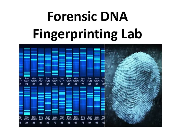

Forensic DNA Fingerprinting Lab
Tools and Technology Used During Lab • p20 micropipette • Restriction enzymes (EcoRI, PstI, HindIII) • Water bath – 37 ° C • Agarose gel electrophoresis • DNA ladder (molecular ruler)
Restriction Enzymes (also known as restriction endonucleases)
What are restriction enzymes? • An enzyme that cuts DNA at specific nucleotide sequences known as restriction sites • Naturally found in bacteria and have evolved to provide a defense mechanism against invading viruses • In bacteria, restriction enzymes selectively cut up foreign DNA in a process called restriction
Restriction Enzymes in Bacteria
Restriction Enzymes • Cut both strands of the DNA double helix • Over 3000 enzymes have been identified • More than 600 available commercially • Routinely used for DNA modification and manipulation in laboratories ( “molecular scissors” )
Restriction Sites • Also known as recognition sites • Generally genetic palindromic sequences • A palindromic sequence in DNA one in which the 5’ to 3’ base pair sequence is identical on both strands • Usually a 4 or 6 base pair sequence
Restriction Sites • The enzymes scan DNA sequences, find a very specific set of nucleotides and make a cut
Ha e III • Ha e III is a restriction enzyme that searches the DNA molecule until it finds this sequence of four nitrogen bases - GGCC 5’ TGACGGGTTCGA GGCC AG 3’ 3’ ACTGCCCAAGGT CCGG TC 5’
Ha e III • Once the recognition site was found Ha e III will go to work cutting (cleaving) the DNA
Blunt Ends versus Sticky Ends • Ha e III produces “blunt ends” when cleaving DNA • Other enzymes produce “sticky ends”
blunt end sticky end
Restriction Enzyme Names • Named after the type of bacteria in which the enzyme is found and the order in which the restriction enzyme was identified and isolated Eco RI for example R strain of E.co li bacteria I as it is was the first E . coli restriction enzyme to be discovered.
Restriction Enzymes and Gene Cloning
Separating Restriction Fragments • Restriction enzymes generate RFLPs (“ rif- lips”) - Restriction fragment length polymorphisms • RFLPs are differences among individuals in the lengths of DNA fragments cut by enzymes • RFLPs can be separated using agarose gel electrophoresis and then analyzed
RFLPs (Restriction Fragment Length Polymorphism)
Separating Restriction Fragments
Gel Electrophoresis • Gel electrophoresis is a laboratory method used to separate mixtures of DNA, RNA, or proteins according to molecular size, shape and charge • The molecules to be separated are pushed by an electrical field through a gel that contains small pores (“molecular strainer”)
Gel Electrophoresis • Larger molecules meet with more resistance when moving through the gel than smaller molecules • Smaller molecules will travel farther than larger molecules
Gel Electrophoresis
More About Gel Electrophoresis • Agarose gel – agarose is a polysaccharide extracted from seaweed • Buffers are used to prepare and cast gels and are also used to fill the electrophoresis chamber • The buffer acts to stabilize pH, maintain molecule shape, and conduct electricity
Nucleic Acid Stains • DNA is not visible • Gels are cast with a nucleic acid stain that, when exposed to UV light, will cause the DNA to fluoresce ( Gel Green ) • As DNA migrates through the agarose gel, the stain will bind to the nucleotides
Forensic Fingerprinting Lab Objectives • Use restriction enzymes to detect differences in the base sequences of individuals • Use gel electrophoresis to analyze DNA samples from suspects and crime scene DNA
DNA Fingerprint
DNA Fingerprinting • Technology used to analyze evidence in law enforcement cases and other applications such as: – Paternity testing – Determine evolutionary relationships among organisms (similarities and differences) – Diagnose genetic disorders or gene testing – Determination of impurities in a sample
Crime Scene DNA Analysis
Paternity Testing
Evolutionary Relationships
Procedure Overview • DNA from crime scene and DNA from 5 potential suspects • Part 1: Restriction Digest of DNA Samples • Part 2: Agarose Gel Electrophoresis of DNA Samples
The Crime and Victim
Suspect 1 – Bobby Joy
Suspect 2 – Paisley Gavin
Suspect 3 – Malcolm Plum
Suspect 4 – Ella Mae Dixon
Suspect 5 – Ruby Warner
The Crime and Victim
Suspect 1 – Angelina Bento
Suspect 2 – Kay McNamara
Suspect 3 – Tori Howard
Suspect 4 – Bella Valentino
Suspect 5 – Abby Farrell
Part I: Restriction Digest of DNA Samples • EcoRI/PstI enzyme mix ( ENZ ) • Crime scene DNA ( CS ) - green • Suspect 1 ( S1 ) - blue • Suspect 2 ( S2 ) - orange • Suspect 3 ( S3 ) - violet • Suspect 4 ( S4 ) - pink • Suspect 5 ( S5 ) – yellow • Ice and a 37 °C water bath
Part II: Agarose Gel Electrophoresis • Add 5 uL of loading dye (LD) into each sample • Load gel as follows: – Lane 1 – S, DNA size standard – 10 µL – Lane 2 – CS, green tube – 20 µL – Lane 3 – S1, blue tube – 20 µL – Lane 4 – S2, orange tube – 20 µL – Lane 5 – S3, violet tube – 20 µL – Lane 6 – S4, red tube – 20 µL – Lane 7 – S5, yellow tube – 20 µL
Recommend
More recommend