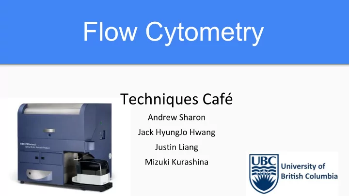

Flow Cytometry Techniques Café Andrew Sharon Jack HyungJo Hwang Justin Liang Mizuki Kurashina
What is Flow Cytometry? ● High-throughput technique to analyse and/or sort cells ● Utilizes microfluidics and lasers to measure parameters
What Is It Used For? Measure many different parameters on the ● same cell Protein expression ○ ○ DNA content Immunophenotype cells ● ● Sorting cells into different tubes based on phenotype (Fluorescence-Activated Cell Sorting: FACS) Determine what phase of the cell cycle a ● cell is in DOI 10.1007/s002530100673
Preparation of flow cytometry Requires 5 operating units ● ○ Light source (mercury lamp or laser) ○ Flow chamber ○ Optical filter ○ Photomultiplier for detection of wavelengths ○ Data processing unit Many cells (100,000+) ● ● Determination of what will be measured (Size, Granularity, Expression of Protein, etc.) Necessary reagents for different conditions ● ○ Size and Granularity: no reagents required Expression of Surface Proteins: Antibodies with conjugated fluorophore against ○ protein of interest, additional secondary antibodies against primary antibodies ○ Expression of Cytoplasmic Proteins: Fixation and pemeabilization of cell (ex. Paraformaldehyde/Saraponin) ○ Expression of Secreted Proteins: Golgi block
How does it work? Sample Data Preparation Processing Hydrodynamic FACS Focusing (optional) Light Laser Detection
Step 1: Hydrodynamic Focusing ● Hydrodynamic focusing allows single cell to move through the laser beam
Step 2: Laser ● Forward scatter (FS) proportional to size of cell detected ● Side scatter (SS) proportional to granularity of cell ● Detect presence of fluorophore via wavelength
Step 3: Detection ● Each cell moves through laser and is individually analyzed ● FS and SS light, and fluorescence are split into defined wavelengths ● Optical filter filters light so that each sensor detects fluorescence at a specific wavelength
Step 4 (optional): FACS ● As cells pass through the laser, the cell is given an electronic charge, depending on the fluorescence of the cell ● Deflection plates attract cells accordingly into collection tubes ● Advantageous because you can purify two or more populations for further research
Step 5: Data Processing ● The amount a cell scatters or fluoresces light is measured and displayed as a histogram eg. allows us to detect abnormal imbalance in CD4+/CD8+ T cells -> to see if HIV has resulted in actual immuno-suppression doi:10.1038/nprot.2009.117
Questions & Answers Thank you for watching our presentation!
Advantages & Disadvantages Advantages Disadvantages Measurements on large number of - Spatial overlap of fluorophores and tedious - optimization of experiments cells eg. characterize Ag expression on cell by - Non-specific binding of antibodies can make cell with large population of cells results difficult to interpret without suitable (important in diagnosis of blood controls disorders - lymphoma, leukemia.. etc) - Requires a suspension of single cells or other - Measured cells can be physically particles, with minimum clumps and debris. sorted for further studies -> This means that the tissue architecture and any information about the spatial relationship between different cells are lost when single cells or nuclei are prepared. -> does not work well for cells that tend to stick together (eg. carcinoma or sarcoma)
Recommend
More recommend