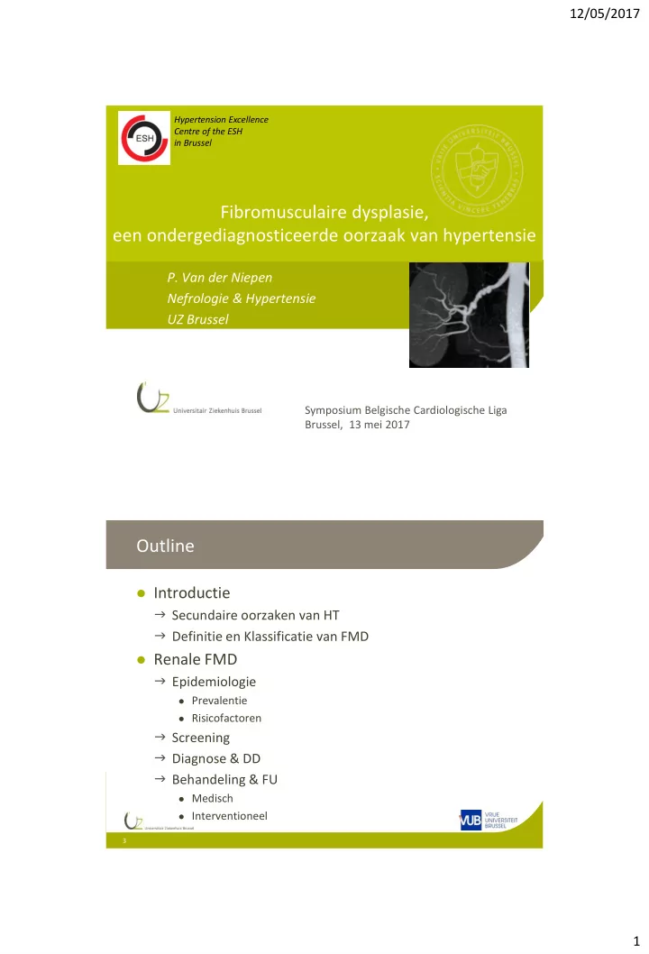

12/05/2017 Hypertension Excellence Centre of the ESH in Brussel Fibromusculaire dysplasie, een ondergediagnosticeerde oorzaak van hypertensie P. Van der Niepen Nefrologie & Hypertensie UZ Brussel Symposium Belgische Cardiologische Liga Brussel, 13 mei 2017 Outline Introductie Secundaire oorzaken van HT Definitie en Klassificatie van FMD Renale FMD Epidemiologie Prevalentie Risicofactoren Screening Diagnose & DD Behandeling & FU Medisch Interventioneel 3 1
12/05/2017 Hypertensie: Etiologie >90% essentieel Cave BD verhogende <10% secundair geneesmiddelen e.a. Renale hypertensie Primaire nierziekte Progressieve nierinsufficiëntie Renovasculaire hypertensie Endocriene oorsprong Primair hyperaldosteronisme (S van Conn) Feochromocytoom Cushing syndroom en ziekte, …. Obstructief SlaapApnoe S, ... ….. 4 11 2
12/05/2017 Introduction Definition & Classification of FMD FMD is an idiopathic, segmental, non- atherosclerotic and non-inflammatory disease of the musculature of arterial walls, leading to stenosis of small and medium-sized arteries. Histopathological classification: three main types: 1. Intimal FMD (5%) ~ Harrison & McCormack (1971) 2. Medial FMD (>85%) • Intimal Fibroplasia (1 - 2%) 3. Perimedial FMD (10%) • Medial FMD (>85%) • Medial fibroplasia (60 – 70%) • Perimedial fibroplasia (15 – 25%) • Medial hyperplasia (5 – 15%) • Adventitial FMD (<1%) 12 Persu et al. J Hypertens 2014; 32(7):1367-78 . O’Connor et al. J Am Heart Assoc 2014; 3: e001259. Introduction Angiographic Classification: 3/ 2 angiographic types Multifocal (‘ string-of- beads’ appearance ), unifocal (solitary stenosis <1 cm in length), and tubular (stenosis 1 cm in length) (Kincaid OW et al. Am J Roentgenol 1968;104:271-82). As the two last categories differ only by the length of the diseased segment, it was proposed to group them under the general term unifocal (Savard S, et al. Circulation 2012; 126:3062 – 69). 80% Medial FMD Intimal FMD 13 Persu et al. Eur J Clin Invest 2012; 42:338-347. Persu et al. J Hypertens 2014; 32(7):1367-78. 3
12/05/2017 Introduction Angiographic Classification: 3/ 2 angiographic types Multifocal (‘ string-of- beads’ appearance ), unifocal (solitary stenosis <1 cm in length), and tubular (stenosis 1 cm in length) (Kincaid OW et al. Am J Roentgenol 1968;104:271-82). As the two last categories differ only by the length of the diseased segment, it was proposed to group them under the general term unifocal (Savard S, et al. Circulation 2012; 126:3062 – 69). 80% Lower female prevalence, more severe and early presentation, higher hypertension cure rate after revascularization. Medial FMD Intimal FMD 14 Persu et al. Eur J Clin Invest 2012; 42:338-347. Persu et al. J Hypertens 2014; 32(7):1367-78. Introduction Definition & Classification of FMD The diagnosis of multifocal FMD can be established when a “string -of- beads” appearance is observed in a medium-sized artery, in the absence of aortic involvement or exposure to vasoconstrictor agents. The diagnosis of unifocal FMD can be established in young patients (usually <40 y), in the absence of atherosclerotic plaque, multiple vascular risk factors, inflammatory syndrome or vascular thickening, and familial or syndromic disease. Persu et al. Eur J Clin Invest 2012; 42:338-347. Persu et al. J Hypertens 2014; 32(7):1367-78. 4
12/05/2017 Differential diagnosis The diagnosis of FMD requires exclusion of Arterial dis. of monogenic origin Inflammatory arterial disease 16 Varennes L et al. Insights Imaging 2015; 6:295-307. Screening for renal FMD (patients with HTN) Expert consensus Age <30 years, especially in women (no family history, no other CV risk factors) Grade 3 (180/110 mmHg) , accelerated or malignant HTN True Resistant HTN (BP target not achieved despite triple therapy at optimal doses including a diuretic) Small kidney without history of uropathy Abdominal bruit without apparent atherosclerosis FMD in at least another vascular territory In individuals aged less than 50 years, screening for FMD may also be considered in milder HTN cases. 19 Persu et al. J Hypertens 2014; 32(7):1367-78 . O’Connor et al. J Am Heart Assoc 2014; 3: e001259 5
12/05/2017 Renal artery FMD - Epidemiology FMD is not so rare! In general population: 0,4% (Plouin et al. Orphanet J Rare Dis 2007; 2:28) Prevalence of FMD in potential kidney donors First author Source Potential donors FMD Cases (%) Cragg, 1989 Universities of Iowa, Minnesota, California San 1862 71 (3.8%) Francisco and Los Angeles, Mayo Clinic 1964-86 Neymark, 2000 University of California san Francisco, 1988-98 716 47 (6.6%) Andreoni, 2002 University of North Carolina, 1995-2001 159 7 (4.4%) Kolettis, 2004 University of Alabama, 1995-2001 1176 66 (5.6%) Blondin, 2010 University of Duesseldorf, 2004 - 2008 101 4 (3,9%) McKenzie, 2013 Mayo Clinic, 2000-2011 2640 68 (2,6%) 263 ( 4.0%) Total 6654 In CORAL trial participants: 5.8% (Hendricks et al. Vasc Med 2014; 19:363-7) Renal artery FMD - Epidemiology FMD is not only a disease of young women! 22 De Groote et al. VASA 2017, 1-8. Olin JW et al. Circulation 2012;125:182-90 6
12/05/2017 Renal FMD in a 65 y man with Coronary Heart Disease Renal artery FMD Pathogenesis and Risk factors Genetic • Autosomal dominant with variable penetrance in 60% of cases based on “clinical symptoms” 1 • 11% prevalence angiographically 2 • PHACTR1 (phosphatase and actin regulator 1) 10 : a first confirmed FMD risk locus Hormonal • No difference in gravidity or parity rates, effect on disease progression 3 • Oral contraceptive pill use? 4,5 Mechanical • Ptosis of the right kidney 6 • Repetitive trauma such as hyperextension and rotation of the neck 6 Mural ischemia • Occlusion of the vasa vasorum 7 • Vasospasm (ergotamines, methysergide) 8 • Tobacco use 9 1 Rushton. Arch Intern Med 1980, 2 Pannier-Moreau. J Hypertens 1997, 3 Stanley. Arch Surg 1975, 4 Sang. Hypertension 1989, 5 Hardy-Godon. J Neuroradiol 1979, 6 Lüscher. Mayo Clin Proc 1987, 7 Sottiurai. J Surg Res 1978, 8 Fievez. Med Hypotheses 1984, 9 Sang. Hypertension 1989. 10 Kiando. PLoS Genetics 2016 26 7
12/05/2017 FMD, a familial disease? ( 104 ) (n, 477 ) 10.6 % Pannier-Moreau I et al. J Hypertens 1997; 15:1797-1801. Olin JW et al. Circulation 2012; 125:3182-90. Screening for hereditary FMD It is recommended to question a patient with FMD about precocious HTN, history of dissection, aneurysm, or history of cerebral haemorrhage among his/her first-degree relatives. In case of a positive answer to at least one of these questions, the patient may inform the respective relative(s) about the possibility of hereditary FMD. Persu et al. J Hypertens 2014; 32(7):1367-78 . O’Connor et al. J Am Heart Assoc 2014; 3: e001259 8
12/05/2017 Savard S et al. Hypertension . 2013;61:1227-32 Less Classical presentations of FMD Renal artery aneurysms/ vascular ectasia • US Registry (n, 447): • Flemish Registry (n, 123): • 17% artery aneurysms • 20% artery aneurysms • 33% in renal artery, • 32% in renal artery • 21 % in carotid artery • 44% in carotid artery • Complications: rare • Rupture • Distal emboli • AV fistula with renal vein Abdominal angio-CT scan: renal artery FMD with RAAs: Left artery: type 1 (saccular) aneurysm (2,5 cm diameter) Right renal artery: type 2 (fusiform) aneurysm (1,3 cm ) 35 Olin. Circulation 2012. De Groote. VASA 2017. Varennes. Insights Imaging 2015; 6:295-307. 9
12/05/2017 Less Classical presentations of FMD Renal artery dissection • US Registry (n, 447): 19,7% arterial dissection (22% in renal artery, 75% in carotid artery) • Flemish Registry (n, 123): 11,4% AD (14% RA, 50% CA) • Lacombe (n, 22 isolated renal artery dissection): 45% FMD as cause - Occur esp. tubular stenosis May cause renal infarction (total occlusion or distal emboli) - flank pain, hematuria a/o rapidly progressive HTN 36 Olin. Circulation 2012. De Groote. VASA 2017. Lacombe. J Vasc Surg 2001;33:385-91. Renal artery FMD Clinical manifestations Hypertension is the most common clinical presentation (Renovascular HT) Variable severity Variable onset Epigastric or flank bruit on physical ex Flank pain < dissection, or aneurysm Renal insufficiency: uncommon RA dissection and Renal infarction ( ) CKD Progression to ESRD: rare 37 Persu et al. J Hypertens 2014; 32(7):1367-78 . O’Connor et al. J Am Heart Assoc 2014; 3: e001259. 10
Recommend
More recommend