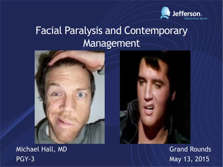

Facial Paralysis and Contemporary Management Michael Hall, MD Grand Rounds PGY-3 May 13, 2015
Overview • Anatomy • General Concepts • Causes • Treatment Options • Static • Dynamic • Management of the… • Brow • Eyelids • Mid-Lower Face • Rehab • Complications and Management of Synkinesis • Future Direction and Research
Anatomy Major Minor
Facial Nerve
General Concepts • Multiple etiologies • Diverse presentation • Not life threatening • Severe QOL implications and psychological impact • Prognosis and outcomes variable
Etiology • Idiopathic • Toxic • Infection • Vascular • Trauma • Neurologic • Iatrogenic • Otologic • Metabolic • Congenital
Grading Facial Nerve Injury
Sunderland Classification Normal
Facial Nerve T esting
Treatment Options • Observation • Conservative • Prednisone: 1mg/kg per day for 7-10 days with slow taper • Acyclovir/Valcyclovir • Chemodenervation, Fillers • Direct Nerve Repair and Cable Grafting • Static Procedures • Facial Sling, Gold Weight • Dynamic Procedures • Nerve, muscle or free tissue transfer
Timing and Considerations with Facial Paralysis • Patient Age • Progression • Complete • Onset • Incomplete • Immediate • Delayed • Patient Expectation • Duration • Status of the Eye • Involved Branches
Primary Nerve Repair and Cable Grafting • Ideal for injuries < 72 hours • Best functional outcomes • Epineural vs Perineural repair • Tension free closure
Cable Grafting • Great Auricular Nerve • 7-10cm • Close proximity • Sural Nerve • 30-75 cm • Several branch points for multiple anastomoses • Medial and Lateral Antebrachial Cutaneous Nerve • Good for concomitant RFFF • ~20 cm harvest
Cable Grafting Pearls • Oblique cut to facilitate grafting more then one branch • Harvest ~25% > defect length • Graft zygomatic and mandibular branches first • At least 6 months for recovery but can be up to 1-2 years • Often times perform static procedures concomitantly
Matteo or Davis
Neural Conduits • End to end anastomosis not possible • Good for gaps < 3cm • Provides support, shape and guidance for axonal regeneration • Limits fibrosis, neuroma formation and FB reaction • No donor site morbidity
Nerve Conduits
Nerve Transfer 1879: Earliest description of nerve transfer by Drobnick, CN XI->VII 1901: Korte performed first CN XII -> VII transfer 1924: Balance recurrent laryngeal -> VII transfer 1971: Scaramella and Smith reported cross facial nerve grafting 1984: Terzis introduced the “babysitter” procedure which combined CFNG and partial XII-VII transfer • Keys to success: Strong contraction, harvest should not result in serious deficit, ability to adhere to rigid rehab program
Cross Facial Nerve Grafting • Contralateral CN VII ideal • <6 months denervation if used as sole procedure • Work medially to laterally to find branches • Map out branches • Ideally match like to like • Make tunnels prior to neurorraphy • Sural Nerve most common • Disadvantages: donor site deficits, long interval of reinnervation, limited donor axons, two coaptation sites, possible sacrifice of function
Scola
Nerve Transfer Options • XII -> VII transfer • Sacrifices ipsilateral XII • Hemiglossal dysfunction, lingual atrophy • End to side, end to end, interposition graft, partial transfer • Babysitter procedure • Partial CN XII transfer with CFNG • Gives immediate motor function while waiting for CFNG reinnervation without ipsilateral tongue paresis
Nerve Transfer Options cont. • CN V transfer • CN XI transfer • Last resort, Mobius syndrome • Less natural result and severe donor site morbidity • Cervical Roots
Static Procedures • Good for temporary paralysis, poor candidates for dynamic procedures, atrophied muscles, failed dynamic procedure • Facial Slings, Gold weight, Lower lid tightening
Static Treatment of the Eyelid • Orbicularis Oculi main depressor of upper lid • Used to treat exposure keratitis • Tarsorrhaphy • Lid Loading (Gold vs Platinum weight) • Lateral Tarsal Strip • Medial/Lateral Canthopexy
Static Treatment of the Brow • Browlift • Direct • Midforehead • Coronal • Trichophytic • Endoscopic
Static Treatment of the Lower Face – Facial Slings Material Advantages Disadvantages Fascia Ease of harvest, autologous Donor site morbidity, tendency of tissue to stretch Lyophilized dermis No donor site, Unpredictable (AlloDerm, ENDURAGen) incorporation into recipient stretching/elongation tissue Expanded Technically easy, local Higher infection and Polytetrafluoroethylne anesthesia, no donor site, extrusion rates (e-PTFE) ease of revision/reversal Multi-vector suture Least invasive, local Unpredictable stretch and suspension anesthesia, quick healing, relaxation, suture breakage easy revision
Facial Slings
Dynamic Muscle Transfer • Restore oral competence • Most commonly used for long standing facial paralysis, restoration of neural input not feasible • Uses functional, innervated and vascular muscle • 2 options • Regional muscle transposition • Free muscle transfer
Regional Muscle Transposition • First description in 1911 by Eden, later popularized in 1977 by Rubin • Most commonly used muscles temporalis, masseter and digastric • Temporalis Muscle Transfer • Innervated by V3, Blood supply deep temporal artery • “Temporal Smile” • Donor site depression, bulge over zygoma, revision surgery, lack of orthodromic muscle contraction
Temporalis Tendon Transfer • Popularized by Labbe • Coronal and nasolabial incisions • Slight overcorrection • Orthodromic muscle contraction, natural vector of pull, less bulky, no donor site depression • 2 versions of technique
Masseter and Digastric Transfer • Masseter • Melolabial incision • Less excursion then temporalis • Digastric • Injury to the marginal branch -> paralysis of the depressor anguli oris and depressor labii inferioris • Anterior belly of digastric • Submental incision
Free Tissue Transfer • Offers possibility of synchronous, mimetic movement • Muscle alone and muscle + soft tissue • Gracilis is first choice and most popular • Located medially and posteriorly to the adductor longus • Attaches to pubic symphysis and medial aspect of tibia • Medial femoral circumflex or profunda femoral artery • Pedicle length ~ 6-8 cm • Anterior branch of obturator nerve
Gracilis Free Tissue Transfer • 2 stage procedure • CFNG • Gracilis FFMT • 1 stage procedure • Masseter nerve • Facial vessels as recipient site • 1cm and 2cm additional length to avoid lip contracture deformity • Early training and muscle stimulation • Complications • Hemorrhage, injury to Stenson’s duct, flap failure, lip contracture, bulkiness, lip asymmetry
Facial Rehab • Nonspecific light massage, electrical stimulation, and repetitions of common facial expressions in a general exercise regimen • Facial Neuromuscular re-education • Enhance desired muscle activity while reducing others • Surface EMG biofeedback and mirror feedback • How to measure success? • Facial Grading System • Facial Disability Index
Management of Synkinesis • Abnormal involuntary movement that occurs simultaneously during voluntary muscle contraction • Aberrant nerve regeneration • Sunderland Class III and above • ~20% patients • Treatments include facial neuromuscular retraining, Botox injection, selective neurolysis or myomectomy
Future Direction and Research • Platelet rich plasma • Neural tube additives • Facial Analysis
Platelet Rich Plasma
Neural T ube Additives
Facial Analysis • Recently, computer analysis has been used to quantitatively measure facial asymmetry and thus synkinesis • Increased sensitivity and reliability • Facial Assessment by Computer Evaluation (FACE) • Peak Motus Motion Measurement System • Automated Facial Image Analysis (AFA) • New focus on 3D analysis
Conclusion • Many options to treat facial paralysis from neurorraphy to free muscle transfer • Onset, timing and duration of paralysis is important • Must match goals of the patient with goals of surgeon • New and dynamic field of facial plastic surgery with continual advancements which will allow for objective data and better results
Thank You • Dr. Heffelfinger • Dr. Krein
Recommend
More recommend