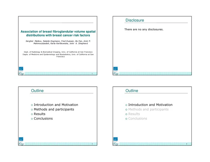

6/8/2017 Disclosure Association of breast fibroglandular volume spatial There are no any disclosures. distributions with breast cancer risk factors Serghei Malkov, Natalie Engmann, Fred Duewer, Bo Fan, Amir P. Mahmoudzadeh, Karla Kerlikowske, John A. Shepherd Dept. of Radiology & Biomedical Imaging, Univ. of California at San Francisco Depts. of Medicine and Epidemiology and Biostatistics, Univ. of California at San Francisco 1 Outline Outline � Introduction and Motivation � Introduction and Motivation � Methods and participants � Methods and participants � Results � Results � Conclusions � Conclusions 3 4 1
6/8/2017 Motivation and purpose Studies on regional breast density and cancer risk Few previous studies have assessed subregional breast � Study motivation density, and suggest that cancers are typically found in the densest local region of the breast tissue (1, 2), usually the upper outer breast quadrant (2). However, Vachon et al. (3) Most studies of breast density to date have examined the did not find evidence that tumor occurrence was higher in the association between density in the whole breast (or global breast upper outer quadrant in a study population containing both density) and breast cancer risk. ductal carcinoma in situ (DCIS) and invasive breast cancers. Breast density varies through the breast with discernible patterns, including regions of peak density and areas of primarily fat. Previous research examining regional breast density has � Although breast density is one the strongest known risk factor primarily used manual segmentation techniques and for breast cancer, little is known about the spatial variation of operator-dependent thresholding methods to calculate the breast density and its relationship to breast cancer risk and risk MLO planar breast density overall and in each region, and spatial factors. autocorrelation statistics including the Moran’s I statistic to From Pereira 2009 measure regional clustering of tissue (1, 2). The purpose of this study was to determine association of breast cancer risk and risk factors with spatial distribution It remains uncertain association of the spatial distribution of of fibroglandular tissue volumes. � breast density with prediction of breast cancer risk. CC In this study, we estimated the effect of common breast cancer risk factors on differences in fibroglandular tissue volumes in 100 � The novel contribution of our study to spatial breast density subregions of the breast. concludes in using accurate volumetric compositional breast Risk factor groups: menopause status, first-degree family history density maps, voxel based morphometry method, and variety of breast cancer (yes vs. no), age at first birth (<30 years vs. of breast cancer risk factors. ≥30 years, and prior history of biopsy, body mass index (BMI: p-value kg/m2) <25 vs. ≥25 categories, race/ethnicity (Caucasian and 1. Pinto Pereira SM et al. 2011 CEBP 20(8):1718 Asian). 2. Pereira SMP et al. 2009 Breast cancer research 11(3):R33 We used the voxel-based morphometry technique which allows 3. Vachon CM et al 2007 Breast Cancer Res 9(6):217 region-by-region group discrimination. From Pereira 2011 5 6 Outline Voxel based morphometry Voxel-based morphometry (VBM) is a visualization � and statistical method in which images are co- registered such that to the same relative position can be statistically compared without regard to object size or distortion. � Introduction and Motivation VBM is widely known for brain studies and there is no � � Methods and participants VBM of brain previous application to breast tissue density. � Results One hundred regions, 8.5x8.5 mm were defined � relative to the nipple, the skin edges, and the chest � Conclusions wall on each breast image. The percent fibroglandular density (%FGV) and � fibroglandular volume (FGV) were estimated for each ROI. � The left and right image ROI values were averaged after mirroring the right breast to the LEFT to keep mediolateral symmetry. Example mammogram showing the 100 regions 7 8 2
6/8/2017 Participants Breast density by Single x-ray absorptiometry For the two-compartment model (0 and 100 refer to fat and fibro-glandular tissue Cases and controls were recruited from an underlying screening � references) and monochromatic case Att can be defined by: mammography cohort at the California Pacific Medical Center I Att ln t t Breast Health Center (CPMC), a participating site of the San = − = + 0 0 100 100 I µ µ 0 Francisco Mammography Registry. The percentage of fibroglandular volume per total breast volume in the pixel at (x,y) location: ( , ) ( , ) Att x y Att x y % ( , ) 0 * 100 Cases were women diagnosed with either invasive breast cancer FGV x y − � ( , ) ( , ) = Att x y Att x y or ductal carcinoma in situ, who had a screening mammogram a 100 0 − year or more before breast cancer diagnosis. Thus, we measure volumetric compositional breast density using two known composition tissue references Att 100 and Att 0. Three controls without breast cancer were randomly selected for � each case and were matched by age, ethnicity, year of screening mammography exam, and mammography system. The study sample consisted of 275 breast cancer cases and 825 � matched controls. Screening film mammograms were digitized with 150 micron � pixel sizes. Breast density and thickness maps Breast image pixel calibration Shepherd et al ., Technol. Cancer Res. Treat. 4 173 (2005) Malkov et al., Medical Physics (2009) 9 9 10 Statistical analysis Outline Covariates: body mass index, menopausal status, family history � of breast cancer, prior history of biopsy, and age at first birth. Statistical comparisons of groups were analyzed by conditional � � Introduction and Motivation logistic regression and generalized linear regression for each ROI. All models were mutually adjusted for covariates. � Methods and participants Principal component analysis (PCA) was applied to transform the � � Results 100 FGV regional measures into orthogonal not correlated variables. � Conclusions Comparison of the principal components vs. global breast � density for breast cancer risk has been performed. 11 12 3
6/8/2017 Group distributions 2D breast voxel based morphometry A:Control A:Premenopausal A:Normal A:Asian B:Case B:Postmenopausal B:Obese B:White Women who developed cancer, on average, had higher fibroglandular volume in all voxels with a maximal differences in the outer lateral and central portion of the breast relative to those women without breast cancer. Women who were post- menopausal had significantly lower breast density in particular regions of the breast with a pattern that suggested a higher density in the periphery of the gland than in the central region. Histogram of fibroglandular volume differences between groups of women from the voxel based morphometry images in previous slides. All 100 regions are shown. Women with BMI>25 had a statistically higher FGV � The largest average difference of group distributions appears to be between the pre difference in the central region and postmenopausal groups. of the breast than those with normal BMI. � Ethnicity status had little average difference but substantial regional differences. White women had a higher FGV in regions near the nipple than � Family history status had no significant spatial differences between regions. Asian women. Voxel-based morphometry of FGV for various groups of women. Only pixels with � Cancer, menopausal, and overweight status groups had both differences in p-values less than 0.0005 are shown in the –log(p-value) images. distributions and spatial significance. 13 14 PCA analysis Correlation PCA1 PCA2 PCA3 PCA4 PCA5 Table 2. Correlation coefficients for demographic variables with the PCA1 seems to capture the outer lateral-medial changes image principal components for control group. of the breast. Risk factors PCA1 PCA2 PCA3 PCA4 PCA5 among PCA2 is an increase in near- controls nipple density relative to the Age -0.41*** 0.05 0.05 0.17*** 0.07 breast interior. Percent Dense 0.68*** -0.27*** -0.3*** -0.15*** 0.08* Area PCA3 demonstrates a difference in density near Percent 0.85 *** -0.25*** -0.13** -0.11** 0.11** the chest wall. Fibroglandular Volume PCA4 captures the variance from medial to lateral Fibroglandular 0.50*** 0.51*** 0.32*** 0.05 0.26*** regions. Volume BMI -0.08 0.35*** 0.25*** 0.18*** 0.08** PCA5 shows the changes in localized central breast Breast Volume -0.13*** 0.62*** 0.36*** 0.12** 0.17*** density. *P<0.05, **P<0.01, ***P<0.0001 � Age was strongly associated with lateral-medial non-uniformity (PCA1 and PCA4). � The highest correlation demonstrates association between PCA1 and %FGV . PCA1-PCA5 for FGV explained 93% of the variance in the FGV . In this image the 5th, � BMI was not associated with PCA1, but was modestly associated with the 50th (mean), and 95th percentile images are shown as well as the difference between other PCA components. the 5th and 95th percentiles. 15 16 4
Recommend
More recommend