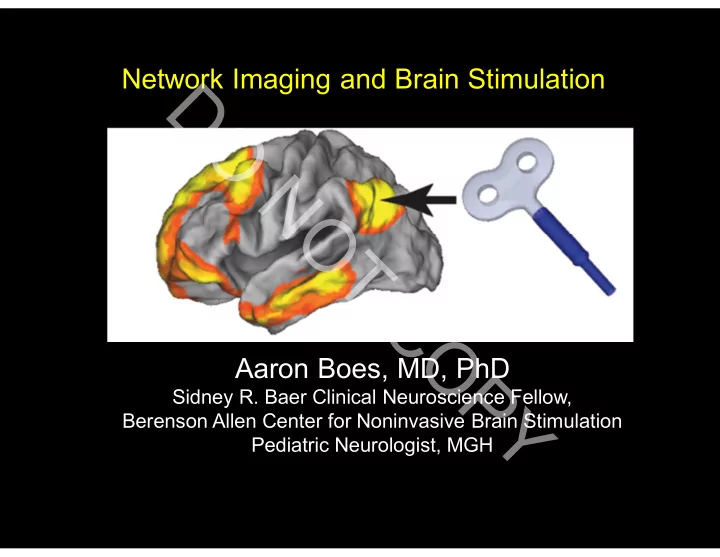

Network Imaging and Brain Stimulation D O N O T C O Aaron Boes, MD, PhD P Sidney R. Baer Clinical Neuroscience Fellow, Y Berenson Allen Center for Noninvasive Brain Stimulation Pediatric Neurologist, MGH
D T opics O 1) A network model for brain function and dysfunction N O 1) What can network imaging contribute to brain T stimulation? C O 2) How does brain stimulation modify networks? P Y
D O N Disclosure: Off-label uses of T MS will be discussed O T C O P Y
D O Mike Fox contributed several slides N O T C O P Y
D O N Please interrupt, ask questions. O T C O P Y
D T opics O 1) A network model for brain function and dysfunction N O 1) What can network imaging contribute to brain T stimulation? C O 2) What can brain stimulation contribute to network imaging? P Y
D Historical Background: Localization of function O N O T C O P Y Paul Broca, 1861
Localization of functional centers >> Network localization of functions D Complex functions arise through interaction among a set of region, each O with specialized processing N O T C O P Y
D Normal functions result O from network-level interactions N Dysfunction also due to O abnormal network activity T C O P Y
D Brain disorders will be treated more effectively O by identifying and targeting therapy toward dysfunctional networks. N O T C O P Y
D T opics O 1) A network model for brain function and dysfunction N O 1) What can network imaging contribute to brain T stimulation? C O 2) What can brain stimulation contribute to network imaging? P Y
D Major types of network imaging O N 1) T ask-based MRI > identify co-active sites O 1) Structural connectivity T - Diffusion tensor imaging C O 2) Correlated functional measures P - BOLD in fMRI, EEG, MEG Y
Classical Neuroimaging Classical functional imaging design D 3 O 2.5 2 % BOLD Change Open Open Open Open N 1.5 O 1 0.5 T Closed Closed Closed Closed 0 0 50 100 150 200 250 C -0.5 T ime (s) -1 O P Open – Closed = Y Slide courtesy of Mike Fox Fox and Raichle (2007) Nat. Rev. Neuro.
BOLD Data Is Very “Noisy” D In task-based fMRI bold signal has “noise” 3 O 2.5 2 % BOLD Change Open Open Open Open N 1.5 O 1 0.5 T Closed Closed Closed Closed 0 0 50 100 150 200 250 C -0.5 T ime (s) -1 O P Open – Closed = Y Slide courtesy of Mike Fox Fox and Raichle (2007) Nat. Rev. Neuro.
BOLD activity oscillates at rest D O N O T C O P Y
BOLD Data Is Very “Noisy” T ask-based activations = 1% of brain energy. D O Majority of energy used to generate % BOLD Change N spontaneous activity, or “fMRI noise” O T Raichle, 2006 C T ime (s) O P Y
Spontaneous Fluctuations (“Noise”) D Using BOLD ‘noise’ as signal in the BOLD Signal O N O T C O P Y
Spontaneous Fluctuations (“Noise”) D Using BOLD ‘noise’ as signal in the BOLD Signal O Left Motor Cortex N 2 Right Motor Cortex O 1.5 T % BOLD Change 1 C 0.5 O 0 P 0 50 100 150 200 250 300 -0.5 Y -1 1 5 9 13 17 21 25 29 33 37 41 45 49 53 57 61 65 69 73 77 81 85 89 93 97 101 105 109 Time (sec) -1.5 Bharat Biswal et al, 1997
Spontaneous Fluctuations are D Specifically Correlated O Left Motor Cortex N 2 Right Motor Cortex O 1.5 T % BOLD Change 1 C 0.5 O 0 P 0 50 100 150 200 250 300 -0.5 Y -1 1 5 9 13 17 21 25 29 33 37 41 45 49 53 57 61 65 69 73 77 81 85 89 93 97 101 105 109 Time (sec) -1.5 Aft er Bhara t Biswal and colleagues (1995) Magne t ic Resonance in Medicine
Generation of Resting State D Functional Connectivity Maps O 2 1.5 N % BOLD Change 1 0.5 O 0 0 50 100 150 200 250 300 -0.5 T -1 Time (sec) C -1.5 O P Y Z score, fixed effects, N = 10 Fox and Raichle (2007) Nat. Rev. Neuro.
D Connectivity of site that is active O during rest, inactive during tasks N Seed Region in Pcc O MPF T 2 C 1.5 O 1 % BOLD Change 0.5 P 0 Y 0 50 100 150 200 250 300 -0.5 -1 -1.5 Time (sec) -2 -2.5
D O N Seed Region in Pcc O MPF T 2 C 1.5 O 1 % BOLD Change 0.5 P 0 Y 0 50 100 150 200 250 300 -0.5 -1 -1.5 Time (sec) -2 -2.5
Default mode network D O N Seed Region in Pcc O MPF T 2 C 1.5 O 1 % BOLD Change 0.5 P 0 Y 0 50 100 150 200 250 300 -0.5 -1 -1.5 Time (sec) -2 -2.5
Regions activated by tasks D Have negative correlation O with default mode network N Seed Region in Pcc O MPF IPS T 2 C 1.5 O 1 % BOLD Change 0.5 P 0 Y 0 50 100 150 200 250 300 -0.5 -1 -1.5 Time (sec) -2 Fox et al. (2005) PNAS -2.5
Regions activated by tasks D Have negative correlation O with default mode network N O T C O P Y Fox et al. (2005) PNAS Fox et al. (2005) PNAS
Resting-state functional connectivity networks correspond to known anatomical paths D O D T I Network Rs-fcMRI Network N O T C O P Y Honey et al. 2009 PNAS
Exponential Popularity of Rs-fcMRI Exponential popularity of rs-fcMRI D O N O T C O P Y Snyder et al. 2012 Neuroimage
Clinical Implications of rs-fcMRI D O • Understanding disease pathophysiology N • Biomarkers / Diagnosis O • Guiding treatment T C O P Y
Clinical Implications of rs-fcMRI D O • Understanding disease pathophysiology N • Biomarkers / Diagnosis O • Guiding treatment T C O P Y
D Sample Case – Using rs-fcMRI to gain insight about O clinical problem N O T C O P Y
D Case O 17 yo girl presents with visual hallucinations N O T C O P Y
D Case O 17 yo girl presents with visual hallucinations N O T - “Eyes were like a zoom lens, going in and out C of focus” O P Y
D Case O 17 yo girl presents with visual hallucinations N O T - “Eyes were like a zoom lens, going in and out C of focus” O - “Scene being drawn in by crayon” P Y
D Case O 17 yo girl presents with visual hallucinations N O T - “Eyes were like a zoom lens, going in and out C of focus” O - “Scene being drawn in by crayon” P - Felt as though she was a referee in a soccer game Y
D Case O 17 yo girl presents with visual hallucinations N O T - “Eyes were like a zoom lens, going in and out C of focus” - “Scene being drawn in by crayon” O - Felt as though she was a referee in a soccer P game Y - Reached for jacket and flowers sprouted from it, then fell over with straight stems, like popsicle sticks
D O N O T C O P Y
D Peduncular Hallucinosis (PH) O N Predominately visual hallucinations following brainstem or thalamus insult O T C O P Y
D Peduncular Hallucinosis (PH) O N Where do lesions causing peduncular hallucinosis localize to? O T C O P Y
D Lesion Overlap Analysis Results O N O T C NN=98 O P Y
Hypothesized mechanism of peduncular hallucinosis D O Subcortical lesions cause a ‘release’ of visual association cortex (Cogan, 1973; Manford, 1998; Kosslyn, 2001; Kazui, 2009) N O T C O P Y Kazui et al, 2009
D O N O T C O P Y
D O N O T C O P Y
Peduncular hallucinosis lesions are anticorrelated to D extrastriate visual cortex O N O T C NN=98 O P Y
D Can this approach be used for other lesion syndromes O that have been challenging to localize? N O T C NN=98 O P Y
D O N O T C O P Y Boes et al, in prep
Conclusion: T he network effects of focal brain lesions can provide insight about the symptoms D O • Networks associated with focal brain lesions N may serve as targets for repetitive T MS to O augment recovery T C O P Y
D O Full description of clinical implications of rs-fcMRI beyond scope of talk N O T C O P Y
Recommend
More recommend