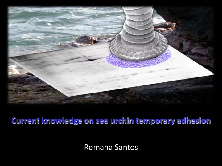

Current knowledge on sea urchin temporary adhesion Romana Santos
My own adhesion network 2006-2009 Pos-doc researcher 2003 Mass Spectrometry Lab ITQB, Portugal Research period Ana Coelho Evolutionary Biomaterials Group Max-Planck, Germany Stanislav Gorb Dagmar Voigt 2009-2013 Assistant researcher 2001-2005 Biomedical and Oral Sciences Research Unit - UICOB PhD thesis Faculty of Dentistry, Portugal Laboratory Marine Biology Manuela Lopes UMH, Belgium Marise Almeida Patrick Flammang 1999-2000 Elise Hennebert Pierre Becker Master thesis Laboratory Marine Biology UMH, Belgium Patrick Flammang
Sea urchin temporary adhesion W1 Chemical characterization and synthesis of adhesives W4 Fabrication of bio-inspired adhesives and their evaluation W2 W3 Structural characterization Mechanical of natural and testing and synthetic theory adhesives
Sea urchin temporary adhesion Structural characterization Sea urchins have: (1) specialized adhesive organs, the oral tube feet mobility resistence stem disc contact adhesion
Sea urchin temporary adhesion Structural characterization Sea urchins have: (1) specialized adhesive organs, the oral tube feet (2) temporary adhesion => attachment / detachment cycles adhesive epidermis
Sea urchin temporary adhesion Structural characterization Sea urchins have: (1) specialized adhesive organs, the oral tube feet (2) temporary adhesion => attachment / detachment cycles adhesive epidermis duo-gland adhesive system
Sea urchin temporary adhesion Structural characterization Sea urchins have: (1) specialized adhesive organs, the oral tube feet (2) temporary adhesion => attachment / detachment cycles ∅ 300-700 nm diversity of adhesive granules adhesive cells ultrastructure released for attachment subtidal DEPTH subtidal-intertidal soft SUBSTRATE hard medium-high low HYDRODYNAMISM duo-gland adhesive system
Sea urchin temporary adhesion Structural characterization Sea urchins have: (1) specialized adhesive organs, the oral tube feet (2) temporary adhesion => attachment / detachment cycles 150x200 nm constant de - adhesive de -adhesive ultrastructure granules cells released for detachment subtidal DEPTH subtidal-intertidal soft SUBSTRATE hard medium-high low HYDRODYNAMISM duo-gland adhesive system
Sea urchin temporary adhesion Structural characterization Sea urchins have: (1) specialized adhesive organs, the oral tube feet (2) temporary adhesion => attachment / detachment cycles (3) adhesive secretion remains on the substrate as a footprint footprint
Sea urchin temporary adhesion Structural characterization Sea urchins have: (1) specialized adhesive organs, the oral tube feet (2) temporary adhesion => attachment / detachment cycles (3) adhesive secretion remains on the substrate as a footprint sponge-like 100-300 nm hydrated in thickness structure
Sea urchin temporary adhesion Mechanical testing Sea urchins have: (1) Tube foot disc tenacities similar to other marine organisms dynamometer Disc tenacity force/area of contact subtrate tube foot disc sea urchin
Sea urchin temporary adhesion Mechanical testing Sea urchins have: (1) Tube foot disc tenacities similar to other marine organisms Sphaerechinus Paracentrotus Echinometra Stomopneustes Colobocentrotus Arbacia Heterocentrotus Taxonomy granularis lividus mathaei variolaris atratus lixula trigonarius Geography Atlantic and Mediterranean Indo-West Pacific Depth subtidal subtidal-intertidal intertidal Substrate soft hard Hydrodynamism very-high low medium-high Tenacity (MPa) 0.20 0.29 0.09 0.22 0.21 0.25 0.54
Sea urchin temporary adhesion Mechanical testing Sea urchins have: (1) Tube foot disc tenacities similar to other marine organisms (2) Tube foot discs perform better on high-energy polar substrates Smooth Tenacity Critical surface Polarity substrates (MPa) area (mJ m -2 ) 0.31 ab Glass 70 0.34 b Polymethylmethacrylate 39 0.17 0.29 ab Polystyrene 33 Polypropylene 0.17 a 32 0.02
Sea urchin temporary adhesion Mechanical testing Sea urchins have: (1) Tube foot disc tenacities similar to other marine organisms (2) Tube foot discs perform better on high-energy polar substrates (3) Tube foot discs replicates the substrate roughness, behaving like a visco-elastic material 2,5 2 Force (mN) 1,5 1 0,5 0 0 20 40 60 80 100 120 Displacement (µm) 2,5 2 Force (mN) 1,5 1 E 1 E 0 0,5 τ 0 0 5 10 15 20 25 30 Time (s)
Sea urchin temporary adhesion Mechanical testing Sea urchins have: (1) Tube foot disc tenacities similar to other marine organisms (2) Tube foot discs perform better on high-energy polar substrates (3) Tube foot discs replicates the substrate roughness, behaving like a visco-elastic material Disc deforms E 1 E 0 Spreads adhesive τ in a thin layer
Sea urchin temporary adhesion Biochemical characterization Sea urchins have: (1) Two sources of adhesive material – extruded and non-extruded Paracentrotus lividus footprints adhesive discs extruded adhesive secretions non-extruded adhesive secretions soluble proteins insoluble proteins complex mixture simpler mixture
Sea urchin temporary adhesion Biochemical characterization Sea urchins have: (1) Two sources of adhesive material – extruded and non-extruded (2) Footprint material is highly insoluble, being composed of at least 13 proteins High concentration of denaturing (2% SDS) and reducing agents (0.5M DTT) => noncovalent (hydrophobic and electrostatic interactions) + disulfide bonds 44% 45% unknown inorganic residues 6 proteins identified by MS – actin, tubulin and histone 3% proteins 2% 6% lipids neutral sugars 7 unidentified proteins - 5 de novo generated sequences with no homology with known proteins
Sea urchin temporary adhesion Biochemical characterization Sea urchins have: (1) Two sources of adhesive material – extruded and non-extruded (2) Footprint material is highly insoluble, being composed of at least 13 proteins
Sea urchin temporary adhesion Biochemical characterization Sea urchins have: (1) Two sources of adhesive material – extruded and non-extruded (2) Footprint material is highly insoluble, being composed of at least 13 proteins 7 unidentified proteins - 5 de novo generated sequences with no homology with known proteins
Sea urchin temporary adhesion Biochemical characterization Sea urchins have: (1) Two sources of adhesive material – extruded and non-extruded (2) Footprint material is highly insoluble, being composed of at least 13 proteins (3) Tube feet discs is more soluble but more complex and contains at least 204 unique proteins Response to Regulation; 38,2% Other; 41,2% stimulus; 5,2% Metabolic process; sliced 40 bands 11,5% Localization; 20,1% Interaction with cells/organisms; 8,4% Cellular process; Developmental 72,3% Immune system Growth ; 3,1% process; 10,5% process; 8,4% 32 fractions/band
Sea urchin temporary adhesion Biochemical characterization Sea urchins have: (1) Two sources of adhesive material – extruded and non-extruded (2) Footprint material is highly insoluble, being composed of at least 13 proteins (3) Tube feet discs is more soluble but more complex and contains at least 204 unique proteins More abundant proteins: Response to Regulation; 38,2% Other; 41,2% stimulus; 5,2% 15% actins => cellular cytoskeleton Metabolic process; 11,5% 11% histones => nucleosome assembly + antibacterial activity 9% ras-related proteins => signal Localization; 20,1% Transduction + vesicle-mediated protein Interaction with transport + regulation of exocytosis cells/organisms; 8,4% Cellular process; Developmental 72,3% Immune system 8% tubulins => microtubule based- Growth ; 3,1% process; 10,5% process; 8,4% movement + protein polymerization
Sea urchin temporary adhesion Biochemical characterization Sea urchins have: (1) Two sources of adhesive material – extruded and non-extruded (2) Footprint material is highly insoluble, being composed of at least 13 proteins (3) Tube feet discs is more soluble but more complex and contains at least 204 unique proteins Other interesting protein groups: Response to Regulation; 38,2% Other; 41,2% stimulus; 5,2% Protein biosynthesis (ribossomal protein, eukaryotic Metabolic process; 11,5% initiation factor, elongation factor 1-alpha and ubiquitin) Protein folding and glycosylation (peptidyl- prolyl cis-trans isomerase; protein disulfide-isomerase, dolichyl-diphosphooligosaccharide protein glycosyltransferase ) Localization; 20,1% Interaction with cells/organisms; 8,4% Cellular process; Developmental 72,3% Immune system Growth ; 3,1% process; 10,5% process; 8,4% adhesive protein secretion
Recommend
More recommend