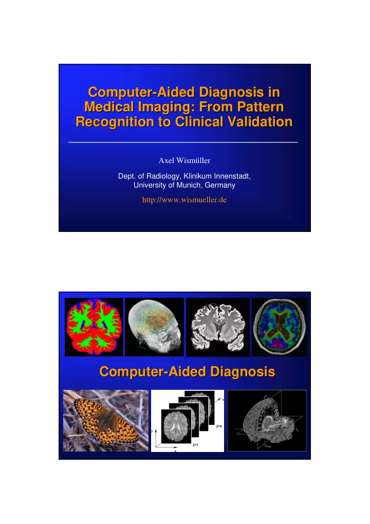

Computer- -Aided Diagnosis in Aided Diagnosis in Computer Medical Imaging: From Pattern Medical Imaging: From Pattern Recognition to Clinical Validation Recognition to Clinical Validation Axel Wismüller Dept. of Radiology, Klinikum Innenstadt, University of Munich, Germany http://www.wismueller.de Axel Wismüller, Dept. of Radiology, University of Munich Computer- Computer -Aided Aided Diagnosis Diagnosis Axel Wismüller, Dept. of Radiology, University of Munich
„Vision“: Computer Vision“: Computer- -Aided Diagnosis Aided Diagnosis „ Image Image Computer Output Medical Expert Diagnosis Axel Wismüller, Dept. of Radiology, University of Munich „Vision“: Computer Vision“: Computer- -Aided Diagnosis Aided Diagnosis „ Glioblastoma multiforme p = 0.95 Fiction ?! ?! Science Fiction Science Midline shift Perifocal edema Axel Wismüller, Dept. of Radiology, University of Munich
Medical Image Image Processing Processing Medical • Segmentation • Registration • Image Sequence Analysis • Classification Axel Wismüller, Dept. of Radiology, University of Munich Medical Image Image Processing Processing Medical Segmentation: Identification of `meaningful´ image components Axel Wismüller, Dept. of Radiology, University of Munich
Medical Image Image Processing Processing Medical Segmentation is the unsolved problem of medical image processing and computer visualization C. Pelizzari Axel Wismüller, Dept. of Radiology, University of Munich Image Segmentation Segmentation Image • Manual segmentation by human experts is time-consuming, expensive • Not feasible in clinical practice → Automatic segmentation is desirable ! Axel Wismüller, Dept. of Radiology, University of Munich
Image Segmentation Segmentation Image Relevant application problem in neuroradiology: Segmentation of 3D MRI data sets of the human brain into the structure classes `Gray Matter´, `White Matter´, and `Cerebrospinal Fluid´ (CSF) Axel Wismüller, Dept. of Radiology, University of Munich Why Brain Brain Segmentation Segmentation? ? Why Potential applications in neurology / psychiatry: • Alzheimer´s Dementia: Identification and monitoring of structural changes by precise volume measurements • Multiple Sclerosis: White Matter Lesions (WML) and quantitative measures for brain atrophy • In general: Clinical studies based on quantitative evaluation of disease progression and therapeutic effects Axel Wismüller, Dept. of Radiology, University of Munich
Motivation: Satellite Satellite Remote Remote Sensing Sensing Motivation: Axel Wismüller, Dept. of Radiology, University of Munich Motivation: Satellite Satellite Remote Remote Sensing Sensing Motivation: Segmentation of multispectral satellite data LANDSAT 6 channels Axel Wismüller, Dept. of Radiology, University of Munich
Multispectral MRI MRI Data Data Sets Sets Multispectral T1- -weighted weighted T2- -weighted weighted T1 T2 Proton Proton Inversion Inversion Density Density Recovery Recovery Axel Wismüller, Dept. of Radiology, University of Munich Multispectral Image Analysis Image Analysis Multispectral • Registration • Pre-segmentation ⇒ Finally: Construction of feature vectors x = (g T1 , g T2 , g PD , g IR ) g j , j ∈{ T1, T2, PD, IR } : signal intensities Axel Wismüller, Dept. of Radiology, University of Munich
Segmentation Approaches Approaches Segmentation • Unsupervised cluster analysis (Vector quantization) Minimal free energy VQ, self-organizing maps, fuzzy c-means, etc. • Supervised classification (GRBF neural network) • Deformable feature map: Mixture between unsupervised and supervised learning component Axel Wismüller, Dept. of Radiology, University of Munich Unsupervised Cluster Analysis Cluster Analysis Unsupervised Assign pixels to codebook vectors according to minimal distance criterion in the feature space... `Gray matter´ `White matter´ `CSF´ Axel Wismüller, Dept. of Radiology, University of Munich
Unsupervised Cluster Analysis Cluster Analysis Unsupervised Combination of all the codebook vectors belonging to a specific tissue class ⇒ Segmentation T1-weighted image Segmentation result Axel Wismüller, Dept. of Radiology, University of Munich GRBF Neural GRBF Neural Network Network Information Flow x ∈ R n Input layer: w j ⎛ ⎞ 2 − ⎜ ⎟ x w − j exp ⎜ ⎟ ρ ⎜ 2 ⎟ 2 ⎝ ⎠ j Hidden layer: = a ( ) x ⎛ ⎞ j − 2 ∑ = ⎜ ⎟ x w N − i exp s j ⎜ ⎟ ρ 2 i 1 2 ⎝ ⎠ i N ∑ = Output layer: ( ) ( ) j a y x s x j Structure = j 1 Axel Wismüller, Dept. of Radiology, University of Munich
Supervised Classification Classification Supervised Acquisition of a training data set: Manual classification of a pixel subset (ca. 1 %) Axel Wismüller, Dept. of Radiology, University of Munich Supervised Classification Classification Supervised Segmentation result of T1-weighted image GRBF classification Axel Wismüller, Dept. of Radiology, University of Munich
Automatic Segmentation Segmentation Automatic Question: Can we re-utilize prevoiously acquired knowledge in order to economize the segmentation procedure? Axel Wismüller, Dept. of Radiology, University of Munich Deformable Feature Feature Map Map Deformable Source Space X Target Space Y Reference data + S + + + + + { } { } μ μ ∈ ν ν ∈ , 1 ,..., q Feature Vectors , 1 ,..., p x y { } { } ∈ ∈ , j 1 ,..., N , j 1 ,..., N w r Test data j j Codebook Vectors μ ν ∈ ∈ n n x , R y , R w r j j A. Wismüller and H. Ritter: The Deformable Feature Map – A Novel Neurocomputing Algorithm for Adaptive Plasticity in Pattern Analysis . Neurocomputing 48:107-139 (2002) A. Wismüller et al.: Fully Automated Biomedical Image Segmentation by Self-Organized Model Adaptiation. Neural Networks 17:1327-1344 (2004) Axel Wismüller, Dept. of Radiology, University of Munich
Deformable Feature Feature Map Map Deformable Source Space X Target Space Y Reference data S + + + + + + { } { } μ ν μ ∈ ν ∈ , 1 ,..., q Feature Vectors , 1 ,..., p x y { } { } ∈ ∈ , j 1 ,..., N , j 1 ,..., N w r Test data j j Codebook Vectors μ ∈ ν ∈ n n x , y , R R w r j j Reference: Test data: Individual Y Individual X Axel Wismüller, Dept. of Radiology, University of Munich System Development Development System Co-registration Training Raw data gray level data, ICC rescaling Gray level PBV Preliminary GRBF inhomogeneity evaluation classification correction Iteration loop GRBF classifi- Vector cation on ICC quantization Cluster assign- Spatial Interpreta- Gray level ment and contingency tion of spectrum of classifi- threshold the CVs the CVs cation Volumes Volumes Spatial Composite cluster WM, GM, WM, GM, contingency CSF, WML CSF, WML assignment maps threshold RC RC Axel Wismüller, Dept. of Radiology, University of Munich
Preprocessing Preprocessing Co-registration Co-registration Training Training Raw data Raw data gray level gray level data, ICC data, ICC rescaling rescaling Gray level Gray level PBV Preliminary GRBF Preliminary GRBF inhomogeneity inhomogeneity evaluation classification classification correction correction Iteration loop Iteration loop GRBF classifi- Vector cation on ICC quantization Cluster assign- Spatial Interpreta- Gray level ment and contingency tion of spectrum of classifi- threshold the CVs the CVs cation Volumes Volumes Spatial Composite cluster WM, GM, WM, GM, contingency CSF, WML CSF, WML assignment maps threshold RC RC Axel Wismüller, Dept. of Radiology, University of Munich Unsupervised Learning Learning Unsupervised Learning Unsupervised Co-registration Co-registration Training Training Raw data Raw data gray level gray level data, ICC data, ICC rescaling rescaling Gray level Gray level PBV Preliminary GRBF Preliminary GRBF inhomogeneity inhomogeneity evaluation classification classification correction correction Iteration loop Iteration loop GRBF classifi- Vector Vector cation on ICC quantization quantization Cluster assign- Cluster assign- Spatial Interpreta- Interpreta- Gray level Gray level ment and ment and contingency tion of tion of spectrum of spectrum of classifi- classifi- threshold the CVs the CVs the CVs the CVs cation cation Volumes Volumes Volumes Spatial Spatial Composite cluster Composite cluster WM, GM, WM, GM, WM, GM, contingency contingency CSF, WML CSF, WML CSF, WML assignment maps assignment maps threshold threshold RC RC RC Axel Wismüller, Dept. of Radiology, University of Munich
Recommend
More recommend