Chapter 5: Treatment Machines for External Beam Radiotherapy Set of - PDF document
Chapter 5: Treatment Machines for External Beam Radiotherapy Set of 126 slides based on the chapter authored by E.B. Podgorsak of the IAEA publication: Radiation Oncology Physics: A Handbook for Teachers and Students Objective: To
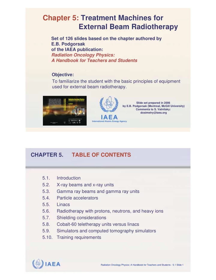
Chapter 5: Treatment Machines for External Beam Radiotherapy Set of 126 slides based on the chapter authored by E.B. Podgorsak of the IAEA publication: Radiation Oncology Physics: A Handbook for Teachers and Students Objective: To familiarize the student with the basic principles of equipment used for external beam radiotherapy. Slide set prepared in 2006 by E.B. Podgorsak (Montreal, McGill University) Comments to S. Vatnitsky: dosimetry@iaea.org IAEA International Atomic Energy Agency CHAPTER 5. TABLE OF CONTENTS 5.1. Introduction 5.2. X-ray beams and x-ray units 5.3. Gamma ray beams and gamma ray units 5.4. Particle accelerators 5.5. Linacs 5.6. Radiotherapy with protons, neutrons, and heavy ions 5.7. Shielding considerations 5.8. Cobalt-60 teletherapy units versus linacs 5.9. Simulators and computed tomography simulators 5.10. Training requirements IAEA Radiation Oncology Physics: A Handbook for Teachers and Students - 5.1 Slide 1
5.1 INTRODUCTION � The study and use of ionizing radiation in medicine started with three important discoveries: • X rays by Wilhelm Roentgen in 1895. • Natural radioactivity by Henri Becquerel in 1896. • Radium-226 by Pierre and Marie Curie in 1898. IAEA Radiation Oncology Physics: A Handbook for Teachers and Students - 5.1 Slide 1 5.1 INTRODUCTION � Immediately upon the discovery of x rays and natural radioactivity, ionizing radiation has played an important role in: • Atomic and nuclear physics from the basic physics point of view. • In medicine providing an impetus for development of radiology and radiotherapy as medical specialties and medical physics as a specialty of physics. • In industry offering many non-destructive measurement techniques and special techniques used in evaluation of oil fields. • In agriculture providing food sterilization and pest control. IAEA Radiation Oncology Physics: A Handbook for Teachers and Students - 5.1 Slide 2
5.1 INTRODUCTION � During the first 50 years of radiation medicine the technological progress was aimed mainly towards: • Development of analog imaging techniques. • Optimization of image quality with concurrent minimization of dose. • Ever increasing energies and beam intensities. � During the past two decades most developments in radiation medicine were related to: • Integration of computers in imaging • Development of digital imaging techniques • Incorporation of computers into therapeutic dose delivery with high energy linear accelerators (linacs). IAEA Radiation Oncology Physics: A Handbook for Teachers and Students - 5.1 Slide 3 5.1 INTRODUCTION � Roentgen discovered x rays in 1895 while experimenting with a Crookes “cold cathode” tube. • Crookes tube is a sealed glass cylinder with two embedded electrodes operated with rarefied gas. • The potential difference between the two electrodes produces discharge in the rarefied gas causing ionization of gas molecules. • Electrons (cathode rays) are accelerated toward the positive electrode producing x rays upon striking it. Photograph of Roentgen’s apparatus IAEA Radiation Oncology Physics: A Handbook for Teachers and Students - 5.1 Slide 4
5.1 INTRODUCTION � Coolidge in 1913 designed a “hot cathode” x ray tube and his design is still in use today. • The main characteristics of the Coolidge tube are its high vacuum and its use of heated filament (cathode). • The heated filament emits electrons through thermionic emission. • X rays are produced in the target (anode) through radiation losses of electrons producing characteristic and bremsstrahlung photons. • The maximum photon energy produced in the target equals the kinetic energy of electrons striking the target. IAEA Radiation Oncology Physics: A Handbook for Teachers and Students - 5.1 Slide 5 5.1 INTRODUCTION � The invention of the cobalt-60 teletherapy machine by Harald E. Johns in Canada in the early 1950s provided a tremendous boost in the quest for higher photon energies and placed the cobalt unit at the forefront of radiotherapy for a number of years. � Most modern cobalt therapy machines are arranged on a gantry so that the source may rotate about a horizontal axis referred to as the machine isocentre axis. � The source-axis distance (SAD) is either 80 cm or 100 cm. IAEA Radiation Oncology Physics: A Handbook for Teachers and Students - 5.1 Slide 6
5.1 INTRODUCTION � Cobalt-60 isocentric teletherapy machine built in the 1970s and 1980s by Atomic Energy of Canada, Ltd. � Source-axis distance = 80 cm IAEA Radiation Oncology Physics: A Handbook for Teachers and Students - 5.1 Slide 7 5.1 INTRODUCTION � At about the same time as cobalt machines clinical linacs were developed. They allowed even higher x-ray energies, eventually eclipsed the cobalt machines and became the most widely used radiation source in modern radiotherapy. � With its compact and efficient design, linac offers excellent versatility for use in radiotherapy through isocentric mounting and provides either electron or x-ray therapy with megavoltage beam energies. IAEA Radiation Oncology Physics: A Handbook for Teachers and Students - 5.1 Slide 8
5.1 INTRODUCTION Standard machines used for modern radiotherapy: � X-ray machine: • Superficial x-ray machine: 50 - 80 kVp • Orthovoltage x-ray machine: 80 - 350 kVp � Cobalt-60 teletherapy machine � Linear accelerator (linac): • Megavoltage x rays: 6 - 25 MV • Electrons: 6 - 30 MeV IAEA Radiation Oncology Physics: A Handbook for Teachers and Students - 5.1 Slide 9 5.1 INTRODUCTION Specialized machines used for modern radiotherapy: � Microtron: megavoltage x rays and electrons � Betatron: megavoltage x rays and electrons � Neutron machines • Neutron generator: (d,t) machine producing 14 MeV neutrons • Cyclotron accelerating protons � Proton machines • Cyclotron • Synchrotron IAEA Radiation Oncology Physics: A Handbook for Teachers and Students - 5.1 Slide 10
5.2 X-RAY BEAMS AND X-RAY UNITS � Clinical x-ray beams typically range in energy between 10 kVp and 50 MV and are produced in x-ray targets when electrons with kinetic energies between 10 keV and 50 MeV strike special metallic targets. � In the target most of the electron’s kinetic energy is transformed into heat, and a small fraction of the kinetic energy is emitted in the form of x ray photons which are divided into two categories: • Characteristic x rays following electron - orbital electron interactions • Bremsstrahlung photons following electron - nucleus interactions IAEA Radiation Oncology Physics: A Handbook for Teachers and Students - 5.2 Slide 1 5.2 X-RAY BEAMS AND X-RAY UNITS 5.2.1 Characteristic x rays � Characteristic X rays result from Coulomb interactions between the incident electron and atomic orbital electrons of the target material (collision loss). � The orbital electron is ejected from its shell and an electron from a higher level shell fills the resulting orbital vacancy. � The energy difference between the two shells is: • Either emitted from the target atom in the form of a photon referred to as characteristic photon. • Or transferred to another orbital electron that is ejected from the target atom as an Auger electron. IAEA Radiation Oncology Physics: A Handbook for Teachers and Students - 5.2.1 Slide 1
5.2 X-RAY BEAMS AND X-RAY UNITS 5.2.1 Characteristic x rays � h � Characteristic photon and Auger electron e KLM following a vacancy in the atomic K shell. Energy of K photon: � ( h � ) K � = ( E B ) K � ( E B ) L � Energy of e KLM Auger electron: ( E K ) e KLM = ( E B ) K � ( E B ) L � ( E B ) M IAEA Radiation Oncology Physics: A Handbook for Teachers and Students - 5.2.1 Slide 2 5.2 X-RAY BEAMS AND X-RAY UNITS 5.2.1 Characteristic x rays � � Fluorescent yield gives the number of fluorescent (characteristic) photons emitted per vacancy in a shell. � K-shell vacancies are the most prominent sources of characteristic x rays. � K � Range of : � K = 0 for small Z • . � K = 0 . 5 for Z = 30 • . � K = 0 . 96 for high Z • . IAEA Radiation Oncology Physics: A Handbook for Teachers and Students - 5.2.1 Slide 3
5.2 X-RAY BEAMS AND X-RAY UNITS 5.2.2 Bremsstrahlung (continuous) x rays � Bremsstrahlung x rays result from Coulomb interactions between the incident electron and the nuclei of the target material. � During the interaction the incident electron is accelerated and loses part of its kinetic energy in the form of brems- strahlung photons. � The interaction is also referred to as radiation loss producing braking radiation. IAEA Radiation Oncology Physics: A Handbook for Teachers and Students - 5.2.2 Slide 1 5.2 X-RAY BEAMS AND X-RAY UNITS 5.2.2 Bremsstrahlung (continuous) x rays � In bremsstrahlung interaction x rays with energies ranging from zero to the kinetic energy of the incident electron may be produced, resulting in a continuous photon spectrum. � The bremsstrahlung spectrum produced in a given x ray target depends upon: • Kinetic energy of the incident electron • Atomic number of the target • Thickness of the target IAEA Radiation Oncology Physics: A Handbook for Teachers and Students - 5.2.2 Slide 2
Recommend


More recommend
Explore More Topics
Stay informed with curated content and fresh updates.