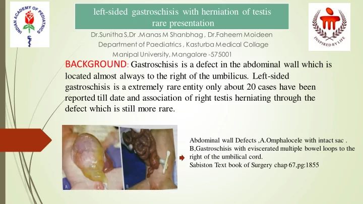

left-sided gastroschisis with herniation of testis rare presentation Dr.Sunitha S,Dr .Manas M Shanbhag , Dr.Faheem Moideen Department of Paediatrics , Kasturba Medical Collage Manipal University, Mangalore -575001 BACKGROUND : Gastroschisis is a defect in the abdominal wall which is located almost always to the right of the umbilicus. Left-sided gastroschisis is a extremely rare entity only about 20 cases have been reported till date and association of right testis herniating through the defect which is still more rare. Abdominal wall Defects ,A.Omphalocele with intact sac . B,Gastroschisis with eviscerated multiple bowel loops to the right of the umbilical cord. Sabiston Text book of Surgery chap 67,pg:1855
Case Presentation We report a 2hr old male neonate who presented with abdominal defect 3 × 3 cm located to the left of umbilicus along with right testis herniation which is an extremely rare entity. Baby also had an associated Ostium secundum Atrial Septal Defect. Neonate born by LSCS to primirigravida mother at 38weeks of gestation, with uneventful antenatal history . Antenatal Ultrasonography done was showing omphalocele. Baby didn’t cry after birth, needing intubation with ventilator support and emergency surgical intervention.
Diagnosis & Treatment Clinical examination, ECHO,NSG to rule out other associations Treatment: Infants born with gastroschisis should be carefully handled to avoid injury to exposed bowel loops and minimize fluid losses. Typically, infants are placed in a warm, saline -filled plastic organ bag up to nipple line. Supportive treatment like IV fluids and IV antibiotics . Surgical Method : In our case primary closure along with right orchidopexy was done.
Discussion Incidents : It is found in 1 among 4,000 live births (1:4,000) THEORIES : Disruption of blood supply to the developing abdominal wall from the omphalomesenteric duct artery by 8 weeks of gestation. Disruption of right vitelline(yolk sac) artery with subsequent body wall damage and gut herniation. Failure of mesoderm to form in the body wall. Rupture of the amnion around the umbilical ring with subsequent herniation of bowel. Abnormal involution of right umbilical vein leading to weakening of the body wall and gut herniation. Abnormal folding of the body wall results in a ventral body wall defect through which gut herniates.[2,4]
Journal of Clinical and Diagnostic Research. 2013 Oct, Vol-7(10): 2300-2302 Conclusion Gastroschisis is a rare entity which is caused to the left of umbilical cord with only 20 cases till now. Gastroschisis with herniation of testis is still rare with only 3 cases recorded till now (table /fig-2).
References 1)Dai H.Chung, Abdominal wall defects: Gastroschisis, Paediatric Surgery , Sabiston Text book of Surgery chap 67:pg 1854-1855, 19 th edition , volume 2. 2)David J. Hackam, Tracy Grikscheit, Kasper Wang, Jeffery S.Upperman, and Henri R.Ford. Paediatric surgery .Schwartz’s PRINCIPLES of SURGERY. Chap 39:pg 1632-1633, 10 th edition . 3)James Wall,MD. Craig T. Albanese , MD. Paediatric Surgery .LANGE CURRENT Diagnosis and Treatment SURGERY Chap 43: pg1259-1260. 14 th edition . 4)Cassandra Kelleher, MD. Jacob C. Langer,MD,Congenital Abdominal Wall Defects Ashcraft’s Paediatric surgery, Chap 48:pg 625 -630, 5 th edition. 5) Aust N Z J Patel R, Eradi B, Ninan G. Mirror-image left-sided gastroschisis. Surg. 2010; 80: 472-73.
Recommend
More recommend