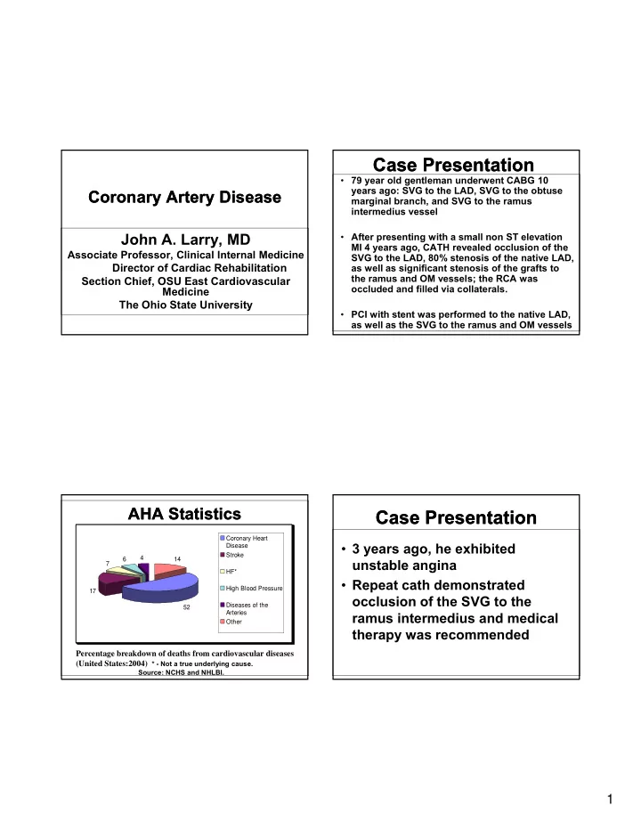

Case Presentation Case Presentation • 79 year old gentleman underwent CABG 10 years ago: SVG to the LAD, SVG to the obtuse Coronary Artery Disease Coronary Artery Disease marginal branch, and SVG to the ramus intermedius vessel John A Larry MD John A. Larry, MD • After presenting with a small non ST elevation MI 4 years ago, CATH revealed occlusion of the Associate Professor, Clinical Internal Medicine SVG to the LAD, 80% stenosis of the native LAD, Director of Cardiac Rehabilitation as well as significant stenosis of the grafts to the ramus and OM vessels; the RCA was Section Chief, OSU East Cardiovascular occluded and filled via collaterals. Medicine The Ohio State University • PCI with stent was performed to the native LAD, as well as the SVG to the ramus and OM vessels AHA Statistics AHA Statistics Case Presentation Case Presentation Coronary Heart • 3 years ago, he exhibited Disease Stroke 4 6 14 unstable angina 7 HF* • Repeat cath demonstrated Repeat cath demonstrated Hi h Bl High Blood Pressure d P 17 occlusion of the SVG to the Diseases of the 52 Arteries ramus intermedius and medical Other therapy was recommended Percentage breakdown of deaths from cardiovascular diseases (United States:2004) * - Not a true underlying cause. Source: NCHS and NHLBI. 1
Case Presentation Case Presentation Case Presentation Case Presentation • Exam • He had been doing well, exercising at a – Pulse 56, BP 138/60 right, 134/60 left, very modest pace 3x a week. resp. rate 16 • 4-5 days prior to office visit, he noted – JVP is normal. No carotid bruits are substernal chest tightness without present present exertional provocation, radiation, or exertional provocation radiation or associated symptoms, lasting 5-10 – Lungs are clear to auscultation and minutes, resolved with a single NTG on 2 percussion occasions. Since that time, he walked – PMI is nondisplaced. S1 and S2 are some, up to 10 minutes at a slow pace normal. A grade 1 systolic ejection without symptoms, and he has exhibited murmur is noted. No gallops or rubs no recurrent chest pain. present Case Presentation Case Presentation Case Presentation Case Presentation • Exam • Current medications include – Abdomen is soft and nontender, with – ASA no organomegaly, aneurysm or bruits – Clopidogrel 75 mg daily – Extremities free of edema, distal pulses are palpable. l l bl – Metoprolol XL 25 mg daily – Isosorbide120 mg daily – Simvastatin 80 mg daily – Lisinopril 10 mg daily – SL NTG 2
Use of Baves theorem to calculate the Use of Baves theorem to calculate the probability of coronary artery disease probability of coronary artery disease mptomatic, HBP, ↑ chol, D.M. typical chest pain 55 y/o M, ty of CAD (%) tic, no risk factors 100 – 80 – (+) ST (+) ST Post-test Probabilit 45 y/o M, asymptomat 45 y/o M, asy 60 – 40 – (-) Exercise ECG (+) Exercise ECG 20 – (-) ST 0 – 0 20 40 60 80 100 Pre-test (Clinical) Probability of CAD (%) JACC 1989; 13: 1653 Prognostic Information in Prognostic Information in Diagnostic studies for evaluation of Diagnostic studies for evaluation of Exercise Treadmill Testing Exercise Treadmill Testing ischemic heart disease ischemic heart disease • Abnormal BP response • Stress EKG • Abnormal Chronotropic Response • Stress ECHO (treadmill or • Impairment in Heart Rate Recovery pharmacologic) • Exercise Duration E i D ti • Stress nuclear (treadmill or St l (t d ill pharmacologic) • Magnitude and Duration of ST Segment Depression • Adenosine/dobutamine MRI • Duke Treadmill Score (Mark, et al Annals Int Med 1987) • Coronary CT angiography � Exercise time on Bruce protocol (mins)- 5x • Cardiac catheterization with coronary maximum ST depression (mm) -4x anginal index angiography (0-no angina, 1 mild angina, 2-limiting angina) 3
Adverse Prognostic Features in Adverse Prognostic Features in Prognostic Data in Stress Testing Prognostic Data in Stress Testing Treadmill/Pharmacologic Nuclear Treadmill/Pharmacologic Nuclear Imaging Imaging • Multiple reversible perfusion defects in 2 or more coronary territories • Quantitatively large myocardial perfusion defects • Transient ischemic dilation of the LV • Lung uptake Circulation. 1998;98:1622-1630 High Risk Features in High Risk Features in Case Presentation Case Presentation Stress/Dobutamine Echo Stress/Dobutamine Echo • Pharmacologic nuclear study • New or worsening wall motion ordered abnormalities in multiple coronary – His typical walking speed limited territories territories – HR independent study • Peak wall motion score index >1.7 – Both issues raised concern a • Drop in LVEF treadmill study would not be adequate 4
Case Presentation Case Presentation Coronary Artery Disease Coronary Artery Disease • Pharmacologic nuclear study ordered – Previous revascularization – By appropriateness criteria published by the ACC/AHA imaging study by the ACC/AHA, imaging study Richard J. Gumina, MD, PhD considered appropriate Associate Professor, Cardiovascular Medicine – As an aside, pharmacologic nuclear Director, Interventional Cardiovascular Research study is preferred in patients with The Ohio State University LBBB or ventricular paced rhythm Case Presentation Case Presentation Coronary Angiogram Video 1 Coronary Angiogram Video 1 Pharmacologic nuclear study findings: Large, moderate to severe reversible perfusion defect in the inferoapical, entire lateral/inferolateral and basal and mid anterior/anterolateral walls, concerning for ischemia. concerning for ischemia No scintigraphic evidence of prior injury. He was referred for left heart catheterization with coronary and graft angiography. 5
Coronary Angiogram Video 2 Coronary Angiogram Video 2 Revascularization Options Revascularization Options • Indications for PCI • Indications for Coronary Artery • Indications for Coronary Artery Bypass Graft Surgery • Hybrid Revascularization Trial Revascularization Options Revascularization Options Coronary Angiogram Video 3 Coronary Angiogram Video 3 Appropriateness Criteria Appropriateness Criteria ACCF/SCAI/STS/AATS/AHA/ASNC 2009 Appropriateness Criteria for Coronary Revascularization A Report by the American College of Cardiology Foundation Appropriateness Criteria Task Force, Society for Cardiovascular Angiography and Interventions, Society of Thoracic Surgeons, American Association for Thoracic Surgery, American Heart Association, and the American Society of Nuclear Cardiology Endorsed by the American Society of Echocardiography, the Heart Failure Society of America, and the Society of Cardiovascular Computed Tomography Manesh R. Patel, MD, Chair, Coronary Revascularization Writing Group, Gregory J. Dehmer, MD, FACC, FACP, FSCAI, FAHA, Coronary Revascularization Writing Group, John W. Hirshfeld, MD, Coronary Revascularization Writing Group , Peter K. Smith, MD, FACC, Coronary Revascularization Writing Group and John A. Spertus, MD, MPH, FACC, Coronary Revascularization February 2009 180 clinical scenarios Appropriateness of revascularization and appropriateness of PCI or CABG individually as the primary mode of revascularization Patel, M. R. et al. J Am Coll Cardiol 2009;53:530-553 6
Appropriateness Criteria: Appropriateness Criteria: Appropriateness Criteria: Appropriateness Criteria: Low-Risk Low-Risk High Risk High Risk • Severe resting left ventricular dysfunction • Low-risk treadmill score ( ≥ 5) (LVEF < 35%) • Normal or small myocardial perfusion • High-risk treadmill score ( ≤ or equal to 11) defect at rest or with stress defect at rest or with stress • Severe exercise left ventricular • Normal stress echocardiographic wall dysfunction (exercise LVEF < 35%) motion or no change of limited resting • Stress-induced multiple perfusion defect wall motion abnormalities during stress (particularly if anterior) • Stress-induced multiple perfusion defects of moderate size Appropriateness Criteria: Appropriateness Criteria: Appropriateness Criteria: Appropriateness Criteria: Intermediate Risk Intermediate Risk High Risk High Risk • Mild/moderate resting left ventricular • Large, fixed perfusion defect with LV dysfunction (LVEF 35-49%) dilation or increased lung uptake • Intermediate-risk treadmill score (-11 to +5) (thallium-201) • Stress induced moderate perfusion defect • Stress-induced moderate perfusion defect • Echocardiographic wall motion without LV dilation or increased lung uptake (thallium-201) abnormality involving > 2segments • Limited stress echocardiographic ischemia developing with low dose dobutamine or with a wall motion abnormality only at at low heart rate (< 120) higher doses of dobutamine involving ≤ 2 segments • Stress echocardiographic evidence of extensive ischemia 7
Recommend
More recommend