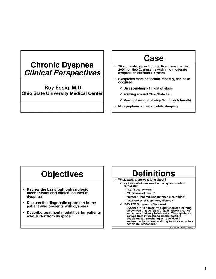
Case Chronic Dyspnea 58 y.o. male, s/p orthotopic liver transplant - PDF document
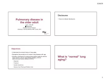
Case Chronic Dyspnea 58 y.o. male, s/p orthotopic liver transplant in 2004 for Hep C, presents with mild-moderate Clinical Perspectives dyspnea on exertion x 5 years Symptoms more noticeable recently, and have occurred: Roy Essig, M.D.
Case Chronic Dyspnea • 58 y.o. male, s/p orthotopic liver transplant in 2004 for Hep C, presents with mild-moderate Clinical Perspectives dyspnea on exertion x 5 years • Symptoms more noticeable recently, and have occurred: Roy Essig, M.D. � On ascending > 1 flight of stairs Ohio State University Medical Center � Walking around Ohio State Fair � Mowing lawn (must stop 3x to catch breath) • No symptoms at rest or while sleeping Definitions Objectives • What, exactly, are we talking about? � Various definitions used in the lay and medical vernacular • Review the basic pathophysiologic • “Can’t get my wind” mechanisms and clinical causes of • “Shortness of breath” dyspnea • “Difficult, labored, uncomfortable breathing” • “Awareness of respiratory distress” • Discuss the diagnostic approach to the � 1999 ATS Consensus Statement patient who presents with dyspnea • Dyspnea is “a subjective experience of breathing discomfort that consists of qualitatively distinct • Describe treatment modalities for patients sensations that vary in intensity. The experience who suffer from dyspnea derives from interactions among multiple physiological, psychological, social, and environmental factors, and may induce secondary behavioral responses.” AJRCCM 1999; 159:322. 1
Epidemiology Mechanisms • Why is this important? • Why is my patient short of breath? � Prevalence in U.S. of diseases that � 2 general components commonly cause dyspnea • Sensation • COPD: 15-30 million – Neural input from a peripheral • Asthma: 15-20 million receptor • Congestive heart failure: 5 million • Perception • Interstitial lung disease: hundreds of – Reaction of the individual to that thousands input � Dyspnea associated with these diseases • Combined, these factors create the has high financial and social cost experience of dyspnea Sensation • Numerous types of afferent input in response to breathing � Chemoreceptors • Central (medulla) Mechanisms of • Peripheral (carotid body) � Mechanoreceptors • Chest wall Dyspnea • Lung parenchyma • Airways • Peripheral muscles � Pulmonary vagal afferents � Irritant receptors • Airways � Direct afferents from central motor output (“Respiratory motor command corollary discharge”) 2
The Carotid Body Perception The sensory organ for respiratory responses to hypoxemia • Patient’s interpretation of and reaction to neural The carotid afferents modified by numerous factors: body senses: � Anxiety � Fear ↓ PaO 2 � Anger and � Depression ↑ PCO 2 � Personality ↓ pH � Previous experiences � Interpersonal relationships • Cause-effect relationships often difficult to sort out E.K. Weir et al. NEJM 2005 Sensation • When afferent impulses from respiratory activity (sensation) does not match outgoing central motor commands, dyspnea occurs Causes of � Hypoxemia � Hypercapnea � Hyperinflation Dyspnea � Increased ventilatory demand � Respiratory muscle weakness � Increased ventilatory impedance • Airways obstruction • Decreased lung compliance • Chest wall abnormalities 3
Causes • What disease might my patient have? � Numerically, most patients with chronic Approach to dyspnea suffer from these 4 diseases: • COPD Dyspnea • Asthma • Interstitial lung disease • Congestive heart failure � The differential, however, includes many others. . . Causes Approach • Pulmonary • Cardiac • How should I evaluate my dyspneic patient? � � Pleural disease Coronary artery disease � Careful history (as always) � � Pulmonary vascular disease Valvular disease • Quality of dyspnea can be helpful � • Pulmonary hypertension Congenital defects � • Thromboembolic disease Arrhythmias – Chest tightness may = bronchospasm � � Neuromuscular disease Pericardial disease – Increased breathing effort often implies � Chest wall abnormalities • Others abnormal mechanical load (e.g., COPD, � Malignancy � Peripheral vascular disease interstitial lung disease, neuromuscular � Large airways obstruction � Deconditioning disease) � Bronchiectasis � Obesity – Sensation of suffocation or air hunger � Vocal cord diseases � often seen in CHF Gastrointestinal diseases � Endocrine/metabolic – Rapid, shallow breathing common in ILD diseases � – “Heavy” breathing often seen in Anemia � deconditioning Psychogenic 4
Approach Approach • History • History � Other � Circumstances of dyspnea • Other medical conditions • Orthopnea often implies CHF, – Risk factors for coronary disease (e.g., HTN, diabetes) diaphragmatic weakness, or GERD – Allergies/atopic disease often seen with asthma • Dyspnea on bending forward often • Medication history suggestive of diaphragmatic limitation (e.g., • Tobacco history diaphragmatic paralysis, obesity) – Significant COPD unlikely in the absence of • Worsening with activity common to most smoking Hx organic causes of dyspnea • Recreational drugs • Presence of dyspnea at rest that improves – Patients often very reticent with this information with exercise strongly suggestive of • Occupational/recreational history psychogenic cause – Environmental lung diseases, chronic infections Approach Approach • History • Exam � Associated symptoms also helpful � HEENT, cardiovascular, lung, and peripheral vascular exam particularly important • Wheezing, chest tightness suggests • Stridor = upper airway obstruction (tumor, bronchospasm tracheostenosis, vocal cord disease) • Dry cough prominent in ILD • Dry/velcro rales often imply ILD • LE swelling suggestive of cardiovascular • “Wet” rales suggestive of CHF disease • Breathing pattern can be useful • Neck tightness, voice change can imply – Accessory muscle use implies increased vocal cord disease workload • Chest pain common in cardiovascular – Rapid, shallow breathing often = ILD disease and pleural disease – “Abdominal” breathing and rib retraction • Recent weight loss or constitutional often = chest wall or neuromuscular disease symptoms can suggest malignancy 5
Case • Medications: cyclosporine, amlodipine, TMP/SMX, pegylated interferon, ribavirin, simvastatin, aspirin, and a multivitamin • Social Hx: never smoker, no alcohol or recreational drugs, former supervisor/office manager in metal industry, but no direct fume/dust exposure, no unusual hobbies; no regular exercise since transplant 5 years prior Case Case • Pt characterized dyspnea as “heavy” breathing with exertion • Vitals: BP 124/86, HR 108, RR 12, afebrile, SpO2 98 % on room air • No changes in dyspnea with position, eating • No symptoms at rest, no nocturnal awakenings • Soft musical 2/6 systolic murmur over precordium, loudest over mitral area; • No associated wheezing, chest tightness/pain, healed chevron incision over upper cough, fevers, weight loss, GERD symptoms, or abdomen; exam otherwise entirely normal swelling • PMH: liver transplant 2004, Hep C, childhood measles, longstanding heart murmur (benign per previous Cardiologist eval) 6
Case Labs • Hemoglobin/hematocrit in most/all patients to evaluate for anemia • Others (metabolic profile, LFT’s, endocrine testing) as history and exam dictation • Our patient: Hgb = 14.1 Assessment High Resolution Chest CT • Chest imaging � Chest X-ray • Reasonable screening tool for gross cardiopulmonary abnormalities � CT chest • More sensitive for subtle parenchymal lung disease, which may be missed on plain x- ray – Particularly important if a diagnosis of interstitial lung disease is being entertained, and should include high- resolution cuts Interstitial Lung Bronchiectasis – Can be done with PE contrast protocol if Disease thromboembolic disease suspected 7
Flow-volume loop in Assessment paradoxical vocal cord motion disorder • Pulmonary function testing � Spirometry • Measure of expiratory airflow • Particularly useful if obstructive lung diseases are suspected (e.g., asthma, COPD) • Can be done before/after bronchodilator to see if obstruction is reversible (e.g., asthma) • Can be done in primary care office • Can (and usually should) be done in conjunction with a flow-volume loop, which gives a visual rendition of expiratory and inspiratory flow • Can be done in concert with inhalational or simulated exercise challenges (for asthma diagnosis) Assessment • Lung volumes � Measurement of capacity of lungs at various moments during the respiratory cycle • Usually abnormally increased in COPD, sometimes in asthma • Usually abnormally decreased in interstitial lung disease • Must be done in pulmonary function lab 8
Assessment Assessment • Maximum inspiratory/expiratory pressure measurement • Diffusing capacity � Indicators of diaphragmatic and accessory � Measure of efficiency of gas exchange in muscle strength, respectively lungs • 6-minute walk test • Decreased in COPD, pulmonary vascular � Ambulation with continuous pulse oximeter disease, anemia, and others for six minutes • Must be done in pulmonary function lab • Both distance and SpO2 recorded • Measure of both exercise tolerance and oxygenation during exercise Full-body Case plethysmography • Spirometry, lung volumes, diffusing capacity: all normal 9
Recommend
More recommend
Explore More Topics
Stay informed with curated content and fresh updates.
