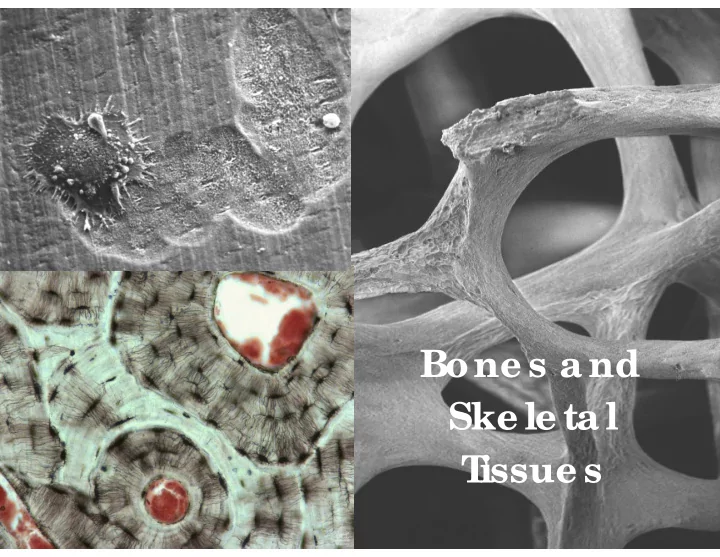

Bone s a nd Ske le ta l T issue s
The Skeleton What are the components of the skeletal system? Cartilage – offers support, resilience and flexibility Hyaline – nose, joint cavities Elastic – ear, epiglottis Fibrocartilage – pubic symphysis & intervertebral discs Bone – organs of the system Also contain nervous, connective and epithelial tissues Ligaments – connect bone to bone Tissue type? Dense regular connective tissue Membranes?
Cartilage What is the importance in relation to the skeletal system? Most bones develop from a cartilage model Provides stability in some freely moving (synovial) joints Provides shock absorption due to ability to compress & expand Cartilage is covered with perichondrium a dense irregular connective tissue to preserve integrity of the cartilage Also aids in growth and repair of the cartilage.
Cartilages in the Adult Body
Growth of Cartilage Two mechanisms for growth: Appositional growth Chondroblasts in surrounding perichondrium produce new cartilage Causes an increase in the width of the cartilage/bone model Interstitial growth Chondrocytes within cartilage divide and secrete new matrix Causes an increase in the length of the cartilage/bone model
Function of Bones Support provides hard framework Movement/Leverage skeletal muscles use bones as levers & along with the muscular system allow for movement of the body Protection of underlying organs Soft tissues of the lungs, and spinal cord, the heart and organs within the pelvic cavity are well protected by bone Storage reservoir for important minerals energy storage in the adipose of yellow marrow Blood-cell formation bone contains red marrow
Classification of Bones Long bones longer than wide containing a shaft plus ends Example: femur Short bones roughly cube-shaped Example: carpals & tarsals Sutural (Wormian) bones Small flat bones found in the sutures of the skull Sesamoid bones Small, round and usually flattened slightly and develop inside of tendons Example: patella Flat bones thin and flattened, usually curved Example: cranial bones (frontal, parietal, temporal, occipital), ribs Irregular bones various shapes, do not fit into other categories Example: vertebrae
Bone Structure Bones are composed of osseous tissue Matrix Cells Outer layer of tissue on bone is the periosteum Inner layer of tissue in bone is the endosteum (lining the marrow cavity)
Microscopic Structure of Bone Matrix Fibers Mainly collagen Ground substance mineralized inorganic material called hydroxyapatite Calcium Phosphate - Ca 3 (PO 4 ) 2 and Calcium Hydroxide - Ca(OH) 2 are the main components that interact to form hydroxyapatite Ca 10 (PO 4 ) 6 (OH) 2 Other substances (calcium carbonate, sodium, magnesium and flouride also become incorporated in the hydroxyapatite providing strength Both are merged in osseous tissue the collagen provides a “framework” for the inorganic salts creating a tissue that is flexible but strong
Microscopic Structure of Bone Cells Osteocytes – the mature bone cell Maintain the matrix by controlling calcium salt deposits in the matrix and the release of calcium into the blood Housed in lacunae that are embedded between the layers of matrix (lamellae) Communicate via canaliculi, allowing osteocyte processes to communicate with adjacent osteocytes across by diffusion or via gap junctions
Microscopic Structure of Bone Cells, cont. Osteoblasts Located on the inner and outer surface of bone Secrete osteoid (organic portion of matrix) which later becomes mineralized Responsible for osteogenesis Once surrounded by matrix it becomes an osteocyte Osteoprogenitor Cells ( progenitor = ancestor ) The mesenchymal cells that differentiate into osteoblasts Found on the inner lining (endosteum) and the outer lining (periosteum) Osteoclasts Large cells that cause osteolysis secretion of acids that dissolve the matrix Osteoclast
Maintenance of the Matrix Matrix maintenance is a balance between osteoclast and osteoblast activity. Osteoclast>osteoblast = bone removal (resporption) Osteoclast<osteoblast = bone addition (deposition) Controlled by hormones that regulate blood Ca 2+ levels Calcitonin (CT) reduces Ca 2+ plasma levels Parathyroid Hormone (PTH) Elevates Ca 2+ plasma levels
Compact vs Spongy Bone The matrix may be highly organized or unorganized Compact bone = organized and relatively solid Spongy bone = unorganized and open design Both types: present in bones have osteocytes, canaliculi and lamellae Compact Bone Functional unit is the Osteon ( Haversian system ) Concentric lamellae surrounding a central canal, collagen fibers spiral in different directions in each layer. Also forms larger circular rings called circumferential lamellae that surround many osteons and the matrix that “fills in” the spaces around the osteons = interstitial lamellae Transverse Section
Compact vs Spongy Bone Spongy Bone Matrix organized into plates of parallel lamellae Forming a lattice called trabeculae Canaliculi open to endosteum to gain nutrients Light weight and high strength Locations of compact & spongy bone: Spongy in the ends of bones and in the marrow or medullary cavities Compact is lining all bones, thickest in areas of high stresses
Microscopic Structure of Compact Bones
Structure of a Typical Long Bone Diaphysis Forms the shaft of a bone Epiphysis Forms the ends of a bone Blood vessels Osseous tissue is well vascularized with vessels in canals in compact bone and running through the trabecular spaces in spongy bone Marrow or Medullary cavity hollow cavity – filled with marrow Membranes Periosteum – continuous with joint capsules and tendons Isolates and protects the bone from surrounding tissue Provides location for nervous and vascular tissue to attach and enter Involved in bone growth and repair (contains osteoprogenitor cells in the inner layer) Sharpey’s fibers (extension of tendons through the periosteum) Endosteum – lines the marrow cavity
Structure of a Long Bone
Structure of Short, Irregular, and Flat Bones Still contains compact bone and spongy No medullary cavity
Bone Design Bone design and stress Anatomy of a bone reflects stresses placed on it Compression and tension forces are largest at external surfaces
Macro to sub-nanostructure of bone
Bone Development Ossification (osteogenesis) – bone-tissue formation Membrane bones – formed directly from mesenchyme by the process of: Intramembranous ossification Clavicle, frontal, parietal occipital, temporal bones Enchchondral bones – develop initially from hyaline cartilage by the process of Endochondral ossification Ossification vs. Calcification Ossification is conversion of cartilage to bone Calcification is the addition of calcium salts to bone
Intramembranous Ossification
Endochondral Ossification
Anatomy of Epiphyseal Growth Areas In epiphyseal plates of growing bones Cartilage is organized for quick, efficient growth Cartilage cells form tall stacks divided into zones Chondroblasts at the top of stacks divide quickly Pushes the epiphysis away from the diaphysis Lengthens entire long bone
Anatomy of Epiphyseal Growth Areas Older chondrocytes signal surrounding matrix to calcify Older chondrocytes then die and disintegrate Leaves long trabeculae (spicules) of calcified cartilage on diaphysis side Trabeculae are partly eroded by osteoclasts Osteoblasts then cover trabeculae with bone tissue Trabeculae finally eaten away from their tips by osteoclasts
Growth of Other Types of Endochondral Bones Short bones – arise from a single ossification center Irregular bones – develop from distinct ossification centers Small long bones Form from a primary ossification center and a single secondary ossification center
Hormonal Regulation of Bone Growth Growth hormone produced by the hypophysis (anterior pituitary gland) Stimulates epiphyseal plates increases rate of division of chondrocytes Thyroid hormone ensures that the skeleton retains proper proportions Sex hormones Promote bone growth Cartilage growth increases, but Rate of osteoblast activity also increases, and Synergistically with hGH and thyroid hormones induces closure of epiphyseal plates (between 18- 25 yrs)
Bone Remodeling Bone deposit and removal Occurs at periosteal and endosteal surfaces Bone remodeling Bone deposition – accomplished by osteoblasts Bone reabsorption – accomplished by osteoclasts
Figure 6.12 Remodeling, Spongy Bone
Repair of Bone Fractures Simple and compound fractures Simple – breaks but does not penetrate skin Compound – breaks and protrudes through skin Treatment by reduction Closed reduction – realignment by hand Open reduction – realignment by surgery
Stages of Healing a Fracture Figure 6.14
Table 6.1 Common Types of Fractures
Table 6.1 Common Types of Fractures
Table 6.1 Common Types of Fractures
Recommend
More recommend