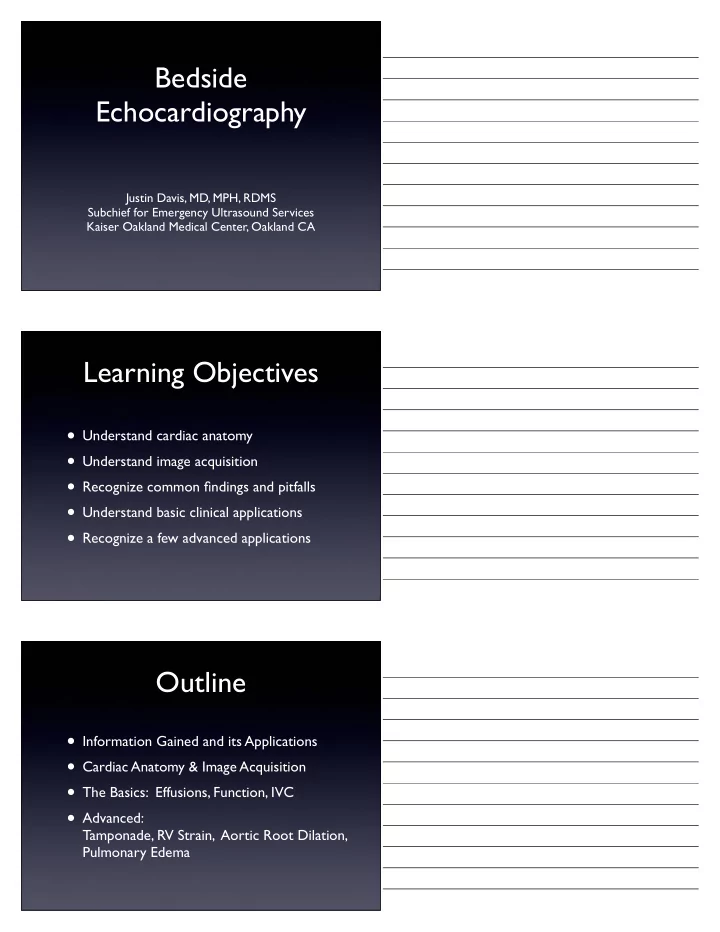

Bedside Echocardiography Justin Davis, MD, MPH, RDMS Subchief for Emergency Ultrasound Services Kaiser Oakland Medical Center, Oakland CA Learning Objectives • Understand cardiac anatomy • Understand image acquisition • Recognize common findings and pitfalls • Understand basic clinical applications • Recognize a few advanced applications Outline • Information Gained and its Applications • Cardiac Anatomy & Image Acquisition • The Basics: Effusions, Function, IVC • Advanced: Tamponade, RV Strain, Aortic Root Dilation, Pulmonary Edema
Information Provided By Bedside Ultrasound The Basics: • Pericardial Effusion • Cardiac Function • Central Venous Pressure Applications Pericardial Cardiac Central Venous Effusion Function Pressure • Trauma • Dyspnea • Cardiac Arrest • Sepsis • Hypotension • Fluid Resuscitation • Chest Pain • Diuresis 4 Echocardiogram Views • Parasternal !! Long Axis • Parasternal !! Short Axis • Apical ! ! ! ! ! 4 Chamber • Subxiphoid !! 4 Chamber
Image Acquisition & Probe Selection • Small footprint • Low frequency Controversy: Probe Orientation General Radiology/EM Cardiology • Indicator • Indicator Screen Screen LEFT RIGHT • Scan from pts • Scan from pts RIGHT LEFT Parasternal Long Axis View (The only one that differs) Moore, C. Current issues with emergency cardiac ultrasound probe and image conventions. Acad Emerg Med 2008; 15: 278-284
Parasternal Long Axis View Probe Indicator Toward right shoulder Parasternal Long Axis View RV LV RV LV Ao Mitral Valve Leaflets DTA Parasternal Short Axis Indicator 90º CCW from Long Axis
Parasternal Short Axis View RV LV Apical 4 Chamber View Indicator similar to Short Axis, Perpendicular plane Apical 4 Chamber View RV LV RA LA
Subxiphoid 4 Chamber View LA LV RA RV Subxiphoid 4 Chamber View Liver RV RA LV LA IVC Indicator toward chin Aim towards thoracic spine
IVC IVC Goals • Assess for IVC fullness • Assess for collapse with spontaneous inspiration • 2-3cm inferior to right atrial junction • Note collapsibility Pitfalls: IVC vs Aorta • Empties into heart ! ! ● Flows deep to heart ! • Flows through liver !! ! ● Flows deep to liver • Undulating Pulsation ! ! ● Bounding Pulsation
Basics: Pericardial Effusions • Anechoic signal (Black) • Between myocardium and pericardium • Effusion should be dependent • Except in trauma or post-op, clinically significant effusions are circumferential Pericardial Effusions False Positive: Fat Pad • Echogenic • Moves with myocardium • Not displaced heart motion • Usually not dependent Pericardial Effusions False Positive: L Pleural Effusion • Only seen posterior/lateral views • In parasternal long axis, extends deep to the descending thoracic aorta (not between DTA and heart) • Use FAST splenorenal view to confirm
False Positive: L Pleural Effusion Pericardial Effusion DTA Pleural Effusion Basics: LV Function • General estimate • Dead to Hyperdynamic • Parasternal long and short axes, look at • Anterior mitral valve leaflet (should come within 1cm of septal wall) • General contraction of LV IVC and CVP IVC Inspiratory CVP Distension collapse Small Complete <5cm H20 Moderate to Full >50% 5-10 Moderate to Full <50% 10-15 Large (>2.5cm) Minimal 15-20cm H20 Large (>2.5cm) None >20cm H20
IVC and CVP • However, don’t need numbers • Give a general estimate • Is the CVP ... extremely low, low, moderate, high, or extremely high? Advanced Finding: Impending Tamponade • 1) In tamponade, intrapericardial pressure restricts atrial filling, therefore IVC WILL (ALMOST ALWAYS) BE DISTENDED • 2) You may see diastolic RA or RV collapse Concave-inward displacement free wall Advanced Finding: RV Strain • Simple explanation: when RV is pushing against high pressure (massive PE) you see: • RV distended and hardly squeezing • LV compressed and under-filled
Normal Parasternal Long Axis LV - Small & Hyperkinetic RV - Large & Hypokinetic Normal Parasternal Short Axis RV - Large & Hypokinetic LV - Small & “D”-Shaped D Hyperkinetic Left Ventricle (Septal Wall Flattening) Advanced Finding Dilated Aortic Root • Ascending aortic dissection often occurs in dilated aortic root • Normal Aortic root < 4cm • Parasternal long axis • Measure 2 cm distal to aortic valve, wall to wall • Neither sensitive nor specific, but may push you along towards the diagnosis
Aortic Root Dilation 5.4cm Parasternal Long Axis Advanced Finding Pulmonary edema • (AKA Alveolar-Interstitial Syndrome) • Lung ultrasound: Same probe, same settings • IVC assesses right-sided congestion, lung assesses left-sided congestion • Scan anterior lung fields • > 3 B-lines per respiratory cycle c/w pulmonary edema B lines Ribs • Arise from the pleural line • Well-defined • Move with lung sliding • Reach the edges of the screen Acute pulmonary edema?
B-lines: The Physics • Fluid-filled interstitium touching the pleural margin • Sound waves go in, and bounce around between air- filled alveoli like a hall of mirrors • Sound waves eventually escape back to the probe CAUTION B-lines are also present in: Cardiogenic pulmonary edema ARDS Pulmonary contusion Pulmonary fibrosis Pneumonia Early atelectasis Pulmonary Edema Applications • Most useful for: • Wheezing vs Cardiac Wheezing • Undifferentiated respiratory failure
Bedside Echo Summary • The Basics: • Significant Pericardial Effusion: Yes/No Circumferential hypoechoic fluid displaced by heart motion • LV Function: Gestalt estimate Note LV contraction and Anterior Mitral Valve leaflet approaching the septum • IVC: Gestalt CVP estimation Note IVC size and collapse with respiration Bedside Echo Summary • Advanced Findings: • Tamponade: Large effusion, plethoric IVC, +/- RA/RV collapse • RV Strain: RV appears enlarged and poorly contracting • Aortic Root Dilation: Parasternal long access, normal <4cm • B-lines of pulmonary edema > 3 per respiratory cycle in bilateral anterior lungs Know there are other causes of B-lines References • Beaulieu Y. Bedside echocardiography in the assessment of the critically ill. Crit Care Med. 2007;35(5 Suppl):S235-49. • Blaivas M. Incidence of pericardial effusion in patients presenting to the emergency department with unexplained dyspnea. Acad Emerg Med. 2001;8(12):1143-1146. • Hernandez C, Shuler K, Hannan H, Sonyika C, Likourezos A, Marshall J. C.A.U.S.E.: Cardiac arrest ultra- sound exam--a better approach to managing patients in primary non-arrhythmogenic cardiac arrest. Resuscitation. 2008;76(2):198-206. • Jones AE, Tayal VS, Kline JA. Focused training of emergency medicine residents in goal-directed echocardiography: a prospective study. Acad Emerg Med. 2003;10(10): 1054-1058. • Jones AE, Tayal VS, Sullivan DM, Kline JA. Randomized, controlled trial of immediate versus delayed goal-directed ultrasound to identify the cause of nontraumatic hypotension in emergency department patients. Crit Care Med. 2004;32(8): 1703-1708. • Lemola K, Yamada E, Jagasia D, Kerber RE. A hand-carried personal ultrasound device for rapid evaluation of left ventricular function: use after limited echo training. Echocardiography. 2003;20(4): 309-312.
Recommend
More recommend