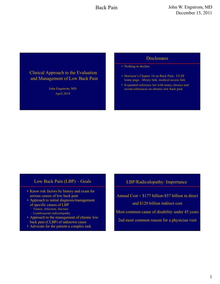

Back Pain John W. Engstrom, MD December 15, 2011 Disclosures • Nothing to declare Clinical Approach to the Evaluation • Harrison’s Chapter 14 on Back Pain: UCSF and Management of Low Back Pain home page; library link; medical access link • Expanded reference list with many classics and John Engstrom, MD recent references on chronic low back pain April 2019 Low Back Pain (LBP) – Goals LBP/Radiculopathy: Importance • Know risk factors by history and exam for serious causes of low back pain Annual Cost = $177 billion-$57 billion in direct • Approach to initial diagnosis/management and $120 billion indirect cost of specific causes of LBP – Tumor, infection, fracture Most common cause of disability under 45 years – Lumbosacral radiculopathy • Approach to the management of chronic low 2nd most common reason for a physician visit back pain (CLBP) of unknown cause • Advocate for the patient-a complex task 1
Back Pain John W. Engstrom, MD December 15, 2011 Acute LBP: Risk Factors for Serious Acute LBP: Risk Factors for Serious Cause - History Cause - Examination Prior history of cancer-strongest correlation Unexplained, documented fever Pain at rest or at night-most common risk missed Unexplained, documented weight loss History of chronic infection-skin, lung, urinary tract Palpation tenderness over spinous processes-C/T/L History of trauma Abdominal, rectal, or pelvic mass Intravenous drug use Patrick’s sign or heel percussion sign Corticosteroid use Straight-leg or reverse straight-leg raising signs History of rapidly progressive neurologic deficit Rapidly progressive focal neurologic deficit Age > 70 years ALBP-Natural History/Treatment LBP – General Examination • 85-90% back to functional baseline in 12 weeks Abdomen-pulsatile mass in 50-75% with AAA • Treat symptoms Spine-palpate spinous processes; use – NSAIDs or acetaminophen for pain paraspinal muscles as a control for non- – Limited bed rest-2 days max; progressive ambulation specific pain – Muscle relaxants if back pain interferes with sleep Hips-Internal/external rotation with leg flexed – Muscle relaxants often not tolerable during a work day Pelvis and Rectum-rare, but don’t forget – Opioids are not a first choice! – Reassurance 2
Back Pain John W. Engstrom, MD December 15, 2011 Initial Approach to Acute Back or Examination Signs Neck Pain Signs that reproduce usual pain symptoms Patrick’s Sign - Hip or buttock pain elicited by Acute LBP 1 internal rotation of the hip with flexion of the leg at the knee Risks for Serious Source? Straight-leg raising – Traction on L5 or S1 roots or sciatic nerve (all posterior to hip) Yes No Reverse straight-leg raising – Traction on L2- Consider infection, tumor, fracture Symptomatic Rx x 3 months No Diagnostic Tests L4 roots or femoral nerve (all ant to hip) 1 Pain < 3 months duration Lumbosacral Radiculopathy - Algorithm 2 -ALBP Suspected Serious Etiology Neurologic Findings Risk factors present Root Motor Reflex Sensory Pain Distribution L4 Quads (knee ext) Knee Medial calf Medial calf Fracture Cancer Infection Rapidly progressive neurologic deficit Leg adduct L5 EHL/EDB/Peronei None Lateral calf, Posterolateral thigh; (foot eversion) dorsal foot Lat calf, dorsal foot ESR, CBC, consider consultation, Plain X-ray/CT Immediate consultation imaging, other lab S1 FDL (toe flexors) Ankle Sole foot Posterior thigh/calf Sole foot 3
Back Pain John W. Engstrom, MD December 15, 2011 Exam for L/S Radiculopathy-Motor L/S Radiculopathy-Sensory • Use smallest bulk muscle avail-most sensitive • L4 - If quad weak, check leg adductors (obturator nerve) • Decreased sensation (negative sensory symptoms) indicates a decrease in sensory function; • L5 -Dorsiflex toes (EDB)/great toe (EHL) • Paresthesias/pain (positive sensory symptoms) -Evert foot (peronei); dorsiflex foot-TA reflect alive nerve cells firing inappropriately • S1 -Toe Flexors-tibial nerve, sciatic nerve • Elicit either a decrease in quantity or quality of • Overcome flexion of toes with fingers-do not screen with sensation (decrease = loss of sensory axons) big toe or foot plantar flexion • Compare light touch from side-to-side • Sensation scale (0 to 10; 0=None, 10 = normal) L/S Radiculopathy-Reflexes L/S Radiculopathy-Sensory Patterns • L4 -Medial calf • Symmetry of the reflex is more important than • L5 -Lateral calf or dorsal foot absolute value (3+ throughout vs. right 3/left 2) • S1- Sole foot • Limbs in analagous positions to compare sides • Sensory loss from peripheral nerve tissue injury • If you can’t get a reflex, then add stretch to the tendon or “reinforcement” root or nerve is in a patch (2Ps: Peripheral-Patch) • CNS tissue injury (spinal cord/brain) produces • L4-sitting or supine, knees bent if supine Circumferential sensory loss in a limb (2Cs: CNS- • L5-No associated reliable reflex Circumferential) • S1-strike Achilles or ball of dorsiflexed foot 4
Back Pain John W. Engstrom, MD December 15, 2011 Lumbar Radiculopathy-Anatomic Diagnosis • Paracentral disk herniation (root in lateral recess) or lateral disk herniation (root in neural foramen) • Bony foraminal stenosis • Tumor, infection, fracture-uncommon • Scarring from prior injury (e.g.-spine surgeries) • Anatomy helps determine etiology of a lumbar radiculopathy and consideration for surgery Natural History of Acute Acute Disk Herniation and Nerve Root Injury: Disk-Related Radiculopathy Compression or Inflammation? • Weber (1983)- If deficit and pain tolerable while waiting, Usually Not Compression spontaneous recovery common - Mobile nerve roots • Saal (1989)-Motor deficits improve with “rehab” and - Nerve roots move during lumbar puncture pain improves over time, but not as fast as with surgery - Gelatinous nucleus pulposus does not compress • Bottom Line: 1) If patient can function with the pain, then the long term outcome is about the same with and - Favorable response to steroids without surgery. 2) If patient’s occupation requires rapid Evidence for Inflammation: Extrusion of nucleus resolution of pain, then seriously consider surgery. pulposis Ò inflammation Ò demyelination 5
Back Pain John W. Engstrom, MD December 15, 2011 Pros of Spine Imaging Radiculopathy is not a Radiologic Diagnosis • May find specific and treatable cause for symptoms • MR Imaging did not replace the neurologist; • Outstanding anatomic definition instead, the imaging findings doubled our work • Non-invasive • “Is the anatomic change clinically significant?” • MRI preferred for anatomy of soft tissues • Imaging establishes anatomic plausibility • CT detects fractures and can define the extent to • Disk abutment to nerve roots often asymptomatic which abnormalities seen on MRI are bony (e.g.- • Does the history, exam, and ancillary testing (as foraminal narrowing from bone vs disk) measures of nerve root function) support the • CT myelography helpful when MRI unavailable anatomic abnormality? Role of Exam and EMG studies Cons of Spine Imaging • Neurologic exam is a qualitative measure of how the nervous system functions (physiology) • Expensive in the U.S. – $143 for spine MRI in Taiwan in 2010! • MRI assesses anatomy of the nervous system – Radiology a profit center or a cost center? • EMG gives a semi-quantitative measure of nerve • Non-specific findings that do not explain the tissue function (physiology) patient’s symptoms are very common • When the anatomy and physiology point to the – May lead to unnecessary additional testing same cause, the probability of a correct diagnosis – May alarm patients and clinicians unnecessarily increases dramatically – Set patient expectations when testing ordered 6
Back Pain John W. Engstrom, MD December 15, 2011 Disk Herniation: Surgical Radiculopathy Diagnostic Stool and Indications the 4 legs: Hx, Exam, Radiol, EMG • History suggests a nerve root injury -Cauda equina syndrome (CES) • Exam shows focal abnormalities suggesting nerve root injury -Spinal cord compression (C/T-spine) • Radiology (MRI) shows anatomic nerve root -Progressive motor weakness by exam injury or compression • EMG, when necessary, establishes nerve root Severe Radicular Pain - Controversial injury and excludes peripheral nerve injury Cauda Equina Syndrome (CES)- CES Diagnosis and Management Symptoms and Signs • Patient describes perineal or perianal numbness • Common etiologies-Herniated disk, tumor, abscess, traumatic displacement of spine • New nocturia or bladder +\- bowel incontinence • Almost never due to chronic spondylotic spinal stenosis • Unable to feel or reduced feeling for toilet paper after urination or bowel movement • Consensus opinion-early surgery (in 1-2 days) better than late; partial syndrome prognosis better than complete • Weakness and numbness in the legs in the distribution of multiple bilateral nerve roots • Send to ER for lumbar spine MRI or CT • Acute, subacute, chronic • Request emergent spine surgery consultation 7
Recommend
More recommend