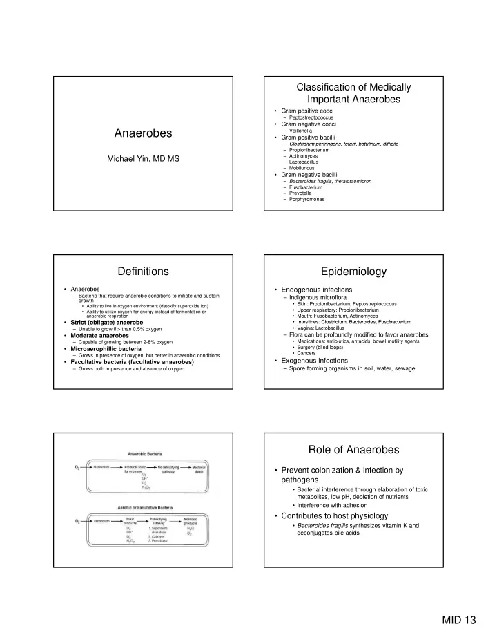

Classification of Medically Important Anaerobes • Gram positive cocci – Peptostreptococcus • Gram negative cocci Anaerobes – Veillonella • Gram positive bacilli – Clostridium perfringens, tetani, botulinum, difficile Clostridium perfringens tetani botulinum difficile – Propionibacterium – Actinomyces Michael Yin, MD MS – Lactobacillus – Mobiluncus • Gram negative bacilli – Bacteroides fragilis, thetaiotaomicron – Fusobacterium – Prevotella – Porphyromonas Definitions Epidemiology • Anaerobes • Endogenous infections – Bacteria that require anaerobic conditions to initiate and sustain – Indigenous microflora growth • Skin: Propionibacterium, Peptostreptococcus • Ability to live in oxygen environment (detoxify superoxide ion) • Upper respiratory: Propionibacterium • Ability to utilize oxygen for energy instead of fermentation or anaerobic respiration • Mouth: Fusobacterium, Actinomyces • Strict (obligate) anaerobe • Strict (obligate) anaerobe • Intestines: Clostridium Bacteroides Fusobacterium • Intestines: Clostridium, Bacteroides, Fusobacterium • Vagina: Lactobacillus – Unable to grow if > than 0.5% oxygen – Flora can be profoundly modified to favor anaerobes • Moderate anaerobes • Medications: antibiotics, antacids, bowel motility agents – Capable of growing between 2-8% oxygen • Surgery (blind loops) • Microaerophillic bacteria • Cancers – Grows in presence of oxygen, but better in anaerobic conditions • Exogenous infections • Facultative bacteria (facultative anaerobes) – Grows both in presence and absence of oxygen – Spore forming organisms in soil, water, sewage Role of Anaerobes • Prevent colonization & infection by pathogens • Bacterial interference through elaboration of toxic metabolites, low pH, depletion of nutrients • Interference with adhesion • Contributes to host physiology • Bacteroides fragilis synthesizes vitamin K and deconjugates bile acids MID 13
Clinical features of anaerobic Virulence factors infections • Attachment and adhesion • The source of infecting micro-organism is – Polysaccharide capsules and pili the endogenous flora of host • Invasion • Alterations of host’s tissues provide – Aerotolerance suitable conditions for development of p • Establishment of infection Establishment of infection opportunist anaerobic infections – Polysaccharide capsule ( B. fragilis ) resists opsonization and phagocytosis • Anaerobic infections are generally – Synergize with aerobes polymicrobial – Spore formation (Clostridium) • Tissue damage • Abscess formation – Elaboration of enzymes, toxins • Exotoxin formation Sites of anaerobic infections Anaerobic cocci • Epidemiology – Normal flora of skin, mouth, intestinal and genitourinary tracts • Pathogenesis – Virulence factors not as well characterized – Opportunistic pathogens, often involved in polymicrobial infections – Brain abscesses, periodontal disease, pneumonias, skin and soft tissue infections, intra-abdominal infections • Peptostreptococcus – P. magnus : chronic bone and joint infections, especially prosthetic joints – P. prevotti and P. anaerobius : female genital tract and intra- abdominal infections • Veillonella – Normal oral flora; isolated from infected human bites Anaerobic gram positive bacilli • No Spore Formation • Spore Formation – Propionibacterium – Clostridium • P. acnes • C. perfringens • C. difficile – Actinomyces • C. tetani C tetani • A. israelii • C. botulinum – Lactobacillus – Mobiluncus MID 13
Propionibacterium Actinomycosis • Cervicofacial • Produces propionic acid as major byproduct of Actinomycosis fermentation – Poor oral hygiene, oral • Colonize skin, conjunctiva, external ear, trauma, invasive dental procedure oropharynx, female GU tract p y – Chronic granulomatous Chronic granulomatous lesions that become • P. acnes suppurative and form sinus – Acne tracts – Slowly evolving, painless • Resides in sebaceous follicles, releases LMW peptide, process stimulates an inflammatory response – Treatment: surgical – Opportunistic infections debridement and prolonged • Prosthetic devices (heart valves, ventricular shunts) penicillin Pilosebaceous follicle Lactobacillus • Facultative or strict anaerobes • Colonize GI and GU tract – Vagina heavily colonized (10 5 /ml) by Lactobacillus crispatus & jensonii – Certain strains produces H 2 O 2 which is bactericidal to Gardnerella vaginalis • Clinical disease – Transient bacteremia from GU source – Bacteremia in immunocompromized host – Endocarditis Actinomyces Case 1 • Facultative or strict anaerobe • 12 year old boy with Acute Myelogenous • Colonize upper respiratory tract, GI, female GU Leukemia (AML) diagnosed 2 mo. ago tract • Pancytopenia after receiving chemotherapy • Actinomycosis • Presented with painful ecchymotic areas on legs Presented with painful ecchymotic areas on legs – Endogenous disease, no person-person spread that rapidly progressed with marked swelling – Low virulence; development of disease when normal and pain over several hours mucosal barriers are disrupted (dental procedure) – Afebrile – Diagnosis made by examination of infected fluid: • Macroscopic colonies of organisms resembling grains of – Crepitus in both legs sand (sulfur granules) – Rapid progression to shock • Culture MID 13
Clostridium perfringens • Epidemiology – GI tract of humans and animals – Type A responsible for most human infections, is widely distributed in soil and water contaminated with feces – Type B-E do not survive in soil but colonize the intestinal tracts of animals and occasionally humans • Pathogenesis g – α -toxin : lecithinase (phospholipase C) that lyses erythrocytes, platelets and endothelial cells resulting in increased vascular permeability and hemolysis – ß-toxin: necrotizing activity – Enterotoxin: binds to brush borders and disrupts small intestinal transport resulting in increased membrane permeability • Clinical manifestations – Self-limited gastroenteritis – Soft tissue infections: cellulitis, fascitis or myonecrosis (gas gangrene) Case 1 • Needle aspirate of ecchymotic area revealed gram- positive bacilli • Blood cultures grew Clostridium perfringens Clostridium Clostridial soft tissue infections • Epidemiology Crepitant cellulitis – Ubiquitous • Present in soil, water, sewage • Normal flora in GI tracts of animals and humans • Pathogenesis – Spore formation • resistant to heat, dessication, and disinfectants • can survive for years in adverse environments – Rapid growth in oxygen deprived, nutritionally enriched environment Fascitis Myonecrosis – Toxin elaboration (histolytic toxins, enterotoxins, neurotoxins) MID 13
Myonecrosis Case 2 • Leukocytosis with 80% neutrophils • Fecal leukocytes • Stool culture neg. for salmonella, shigella l ll hi ll campylobacter, Yersinia spp • Colonoscopy – White plaques of fibrin, mucous and inflammatory cells Clostridial myonecrosis Clostridium difficile • Epidemiology • Clinical course – Endogenous infection – Symptoms begin 1-4 days after inoculation and • Colonizes GI tract in 5% healthy individuals progresses rapidly to extensive muscle necrosis and • Antibiotic exposure associated with overgrowth of C. difficile shock – Cephalosporins, clindamycin, ampicllin/amoxicillin • Other contributing factors: agents altering GI motility, surgery, age, – Local area with marked pain, swelling, oca a ea a ed pa , s e g, underlying illness underlying illness serosanguinous discharge, bullae, slight crepitance – Exogenous infection • Spores detected in hospital rooms of infected patients – May be associated with increased CPK • Pathogenesis • Treatment – Enterotoxin (toxin A) – Surgical debridement • produces chemotaxis, induces cytokine production and hypersecretion of fluid, development of hemorrhagic necrosis – Antibiotics – Cytotoxin (toxin B) – Hyperbaric oxygen • Induces polymerization of actin with loss of cellular cytoskeleton Case 2 C. difficile colitis • Clinical syndromes • 80 year old woman who was treated for a – Asymptomatic colonization pneumonia with a cephalosporin – Antibiotic-associated diarrhea – Pseudomembranous colitis – Well upon discharge from hospital • Diagnosis – Isolation of toxin Isolation of toxin – 10 days later develops multiple, watery loose 10 days later develops multiple watery loose – Culture stools and abdominal cramps • Treatment – Fever, bloody stools, worsened abdominal – Discontinue antibiotics – Metronidazole or oral vancomycin pain – Pooled human IVIG for severe disease – Probiotics (saccharomyces boulardii) – New drugs (nitazoxanide, tolevamer) – Relapse in 20-30% (spores are resistant) MID 13
Recommend
More recommend