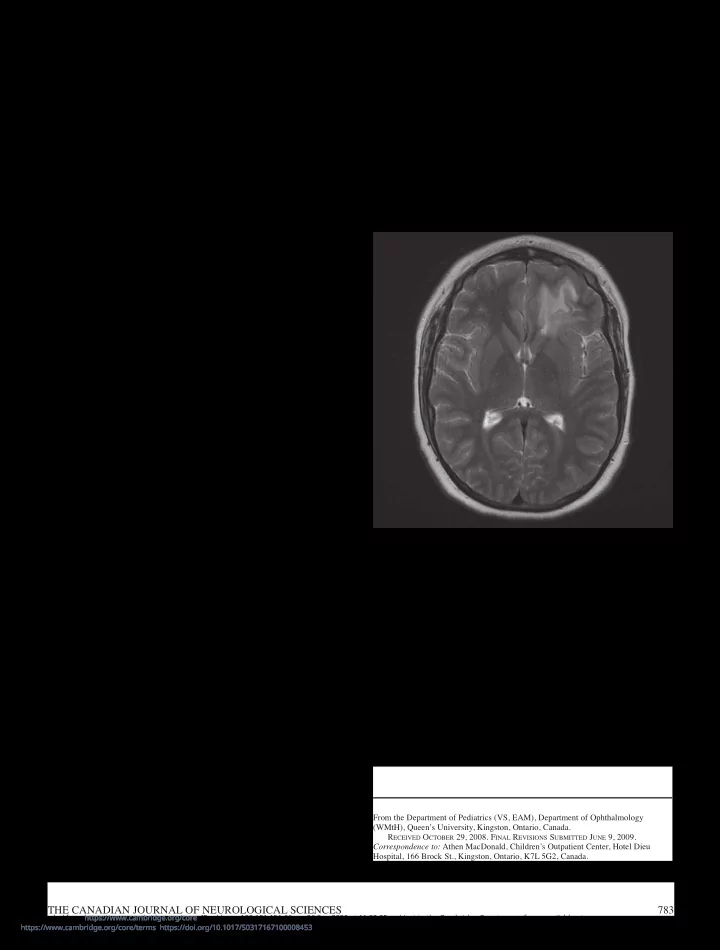

BRIEF COMMUNICATIONS An Unusual Presentation of Neurosarcoidosis in an 11-Year-Old Boy V. Scholten, W.M. ten Hove, E.A. Macdonald Can. J. Neurol. Sci. 2009; 36: 783-786 Sarcoidosis is a chronic, multisystem, granulomatous disease which typically develops between the ages of 20 and 40 years 1 . Neurosarcoidosis occurs in only 5-15% of adults with sarcoidosis, and is seldom reported in children 2-5 . Of the cases described in the literature, children were found more likely to present with seizures, and less commonly space-occupying lesions 6 . The tumefactive brain lesions are difficult to distinguish from the tumefactive demyelinating lesions of multiple sclerosis, from brain neoplasms, acute disseminated encephalo-myelitis (ADEM), or from parasitic foci 7,8 . We report the clinical and radiographic features of an 11-year-old boy with biopsy-proven neurosarcoidosis in order to highlight key features that distinguish neurosarcoidosis from tumefactive demyelination. C ASE REPORT A previously healthy 11-year-old male presented in status epilepticus. The seizure started with an altered level of awareness and focal twitching of the right side of his face. The ictus lasted 45 minutes and responded to intravenous administration of lorazepam and phenytoin. Investigations revealed a peripheral white blood cell count of 28.2 (4.5-13 X10 9 /L), and normal hemoglobin and platelets. Electrolytes, glucose, renal and liver function, and coagulation Figure 1: MR image of the head showing high T2/FLAIR signal parameters were normal. A toxicology screen was negative. involving the inferior left frontal lobe, predominantly within the white matter. Bacterial, viral, and fungal cultures were negative in blood. Cerebrospinal fluid (CSF) analyses revealed 20 X 10 6 /L leucocytes (90% lymphocytes), normal glucose and protein, no malignant cells, and negative bacterial and viral cultures. Polymerase chain reactivity for Herpes viruses was negative. deferred. Phenytoin was weaned and carbamazepine was Oligoclonal banding was detected, but as serum electrophoresis substituted as the primary anti-epileptic drug prior to discharge was not performed, this finding could not be evaluated. Serum from hospital in stable condition. anti-nuclear factor was negative. The boy’s past medical history was unremarkable. He was a Computerized tomography of the head showed a focal, non- good student with appropriate social skills. Family history was enhancing mass of the left frontal lobe with no evidence of mass unremarkable. There was no history of recent travel. effect or hydrocephalus. As shown in Figure 1, magnetic The patient was enrolled in the Prospective Study of the resonance imaging (MRI) demonstrated a region of increased Clinical Epidemiology, Pathobiology and Neuroimaging T2/FLAIR signal involving the white matter of the left frontal Features of Canadian Children with Clinically Isolated lobe with leptomeningeal enhancement after gadolinium contrast Demyelinating Syndromes 9 , which required regular follow-up administration. Magnetic resonance spectroscopy (MRS) demonstrated a lactate peak with normal choline and N- acetylaspartate. A second MRI obtained one week later showed improvement, with normalization of mass spectroscopy. Following recovery from the post-ictal period, the child was noted to have a normal neurological and general physical From the Department of Pediatrics (VS, EAM), Department of Ophthalmology (WMtH), Queen’s University, Kingston, Ontario, Canada. examination, including normal cognition and no focal deficits. R ECEIVED O CTOBER 29, 2008. F INAL R EVISIONS S UBMITTED J UNE 9, 2009. Given the normal examination, and repeat MRI showing Correspondence to: Athen MacDonald, Children’s Outpatient Center, Hotel Dieu improvement in the lesion appearance, brain biopsy was Hospital, 166 Brock St., Kingston, Ontario, K7L 5G2, Canada. THE CANADIAN JOURNAL OF NEUROLOGICAL SCIENCES 783 Downloaded from https://www.cambridge.org/core. IP address: 192.151.151.66, on 26 Sep 2020 at 11:30:25, subject to the Cambridge Core terms of use, available at https://www.cambridge.org/core/terms. https://doi.org/10.1017/S0317167100008453
THE CANADIAN JOURNAL OF NEUROLOGICAL SCIENCES Figure 2: MR image of the head showing re-occurrence of extensive periventricular and subcortical white matter T2 signal abnormality within the inferior left frontal lobe. Figure 3: MR image of the head showing complete resolution of parenchymal signal abnormality within the left frontal lobe, with a new dural thickening and enhancement involving the inferior left frontal skull base. There is also a proptotic left globe, with new left orbital enhancing MRI scans. At three and seven months of follow-up, MRI scans inflammatory change, in continuity with the intracranial left frontal were normal. He complained intermittently of headaches and dural disease. was started on propranolol. The MRI scan at twelve months showed re-occurrence of the left frontal white matter lesion without clinical symptoms (Figure 2). At his 15-month visit the MRI lesion had again resolved. Seventeen months after his initial presentation, the patient noncaseating non-infectious lesions with surrounding complained of worsening headaches. He had insidiously lymphocytes 10 . Meningeal and conjunctival biopsies, the latter developed left ptosis with diplopia on extreme leftward gaze. An often performed blindly, are the most common means of MRI done at this time showed continued resolution of frontal confirming a histological diagnosis 11 . lobe abnormalities, but a new dural thickening and enhancement Involvement of the central nervous system occurs in only 5- of the inferior left frontal skull base and left orbit (Figure 3). The 15% of cases 3,4 . Isolated neurosarcoidosis is rare: Spencer et al 12 ptosis resolved, but he subsequently developed an inflamed found 10-17% of patients had isolated CNS involvement, while conjunctival nodule in the left eye (Figure 4). A conjunctival others detected systemic sarcoidosis in more than 95% of cases biopsy revealed non-necrotizing granulomatous inflammation of sarcoidosis initially presenting with neurological symptoms 10 . and macrophage infiltration, consistent with the diagnosis of Manifestations of neurosarcoidosis include cranial nerve palsies, sarcoidosis (Figure 5). A second attempt at a CSF sample was meningeal involvement, brain lesions, seizures, hypothalamic unsuccessful. Serum angiotensin converting enzyme (ACE) was and endocrine dysfunction, and peripheral neuropathy 13 . Unlike normal and a chest radiograph did not show any granulomatous adults, who characteristically present with a seventh cranial lesions. nerve palsy, prepubertal children are more likely to present with The patient was started on systemic and topical ophthalmic seizures and are perhaps more likely to have a space-occupying steroids which led to resolution of his eye findings. There has lesion 6 . Twenty nine cases of childhood neurosarcoidosis have been no further clinical symptomatology. A recent MRI scan, been reported, with 38% (11/29) presenting with seizures and performed twenty months from initial presentation, showed 24% (7/29) with mass lesions or focal edema on imaging. Of the resolved meningeal enhancement. seven that presented with mass lesions or focal edema on seizures 6 . imaging, three patients presented with D ISCUSSION Neurosarcoidosis may remit spontaneously 7 . Tumefactive demyelinating lesions occur as large solitary Sarcoidosis is a multisystem granulomatous disease of mass lesions or as large focal areas of demyelination of white unknown etiology without an inciting organism identified. matter surrounded by ring enhancement, with little mass effect 14 . Diagnosis requires biopsy confirmation of noncaseating These demyelinating plaques may occur in patients presenting granulomatous inflammation, and histological evidence of with the first attack of multiple sclerosis, in children or adults epithelioid differentiation of macrophages in the center of 784 Downloaded from https://www.cambridge.org/core. IP address: 192.151.151.66, on 26 Sep 2020 at 11:30:25, subject to the Cambridge Core terms of use, available at https://www.cambridge.org/core/terms. https://doi.org/10.1017/S0317167100008453
Recommend
More recommend