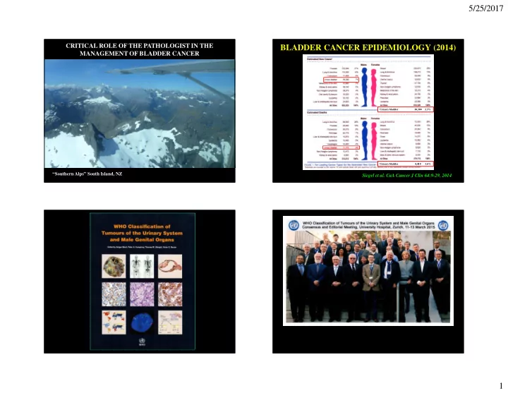

5/25/2017 CRITICAL ROLE OF THE PATHOLOGIST IN THE BLADDER CANCER EPIDEMIOLOGY (2014) MANAGEMENT OF BLADDER CANCER Urinary bladder 18,300 2.3% Urinary bladder 4,410 1.6% “Southern Alps” South Island, NZ Siegel et al. CaA Cancer J Clin 64:9-29, 2014 1
5/25/2017 EORTC RISK TABLES Patients presenting with Ta/Tis/T1 tumors RECURRENCE PROGRESSION* • 6 - Number of tumors • 6 - CIS • 4 - Prior recurrence • 5 - Grade • 3 - Tumor size • 4 - T-category • 2 - Grade • 3 - Number of tumors • 1 - T-category • 3 - Tumor size • 1 - CIS • 2 - Prior recurrence *Progression = development of muscle invasive disease Sylvester et al. Eur Urol 49:466, 2006 NORMAL UROTHELIUM WHO/ISUP 2004/ 2 016 CLASSIFICATION • NORMAL • FLAT LESIONS WITH ATYPIA – Reactive (inflammatory) atypia – Atypia of unknown significance – Dysplasia (low grade intraurothelial neoplasia) – Carcinoma in situ (high grade intraurothelial neoplasia) • PAPILLARY NEOPLASMS – Papilloma – Inverted papilloma – Papillary neoplasm of low malignant potential – Papillary carcinoma, low grade – Papillary carcinoma, high grade • INVASIVE NEOPLASMS Cytokeratin 20 2
5/25/2017 REACTIVE ATYPIA REACTIVE ATYPIA DYSPLASIA DYSPLASIA 3
5/25/2017 CIS “flat urothelial lesion…containing cytologically malignant cells” DYSPLASIA Or Incipient Papillary Neoplasia? Cytokeratin 20 CIS - LARGE CELL CIS - LARGE CELL 4
5/25/2017 CARCINOMA IN SITU CIS – “SMALL CELL” Abundant eosinophilic cytoplasm CIS – DENUDING – VON BRUNN’S NESTS CIS - DENUDING 5
5/25/2017 CARCINOMA IN SITU CARCINOMA IN SITU Pagetoid growth No surface epithelium SV: CIS - PAGETOID CARCINOMA IN SITU Early invasion 6
5/25/2017 CARCINOMA IN SITU – p53 IHC CARCINOMA IN SITU ~ 80% Positive CK20 CARCINOMA IN SITU CARCINOMA IN SITU CK20 AMACR P53 CK 20 7
5/25/2017 REACTIVE ATYPIA UROTHELIAL CARCINOMA IN SITU - LONG TERM OUTCOME SURVIVAL-TYPE 10-Year 15-Year Progression-free 63% 59% Cancer-specific 79% 74% CK20 All-cause 55% 40% p53 Cheng et al, Cancer 85:2469, 2000 CLASSIFICATION AND GRADING OF UROTHELIAL PAPILLOMA PAPILLARY UROTHELIAL NEOPLASMS • Most are small BASED ON TWO KEY FEATURES • Essentially normal • Architectural disruption urothelium • Degree of cytologic atypia • Often vacuolated umbrella cells 8
5/25/2017 PAPILLARY UROTHELIAL NEOPLASM OF PAPILLOMA LOW MALIGNANT POTENTIAL PUNLMP LOW GRADE PUNLMP 9
5/25/2017 LOW GRADE HIGH GRADE LOW GRADE HIGH GRADE HIGH GRADE 10
5/25/2017 HIGH GRADE PAPILLARY BLADDER – PAPILLARY UC 52/164 (32%) papillary UC were grade heterogeneous Cheng et al. Cancer 88:1663-1670, 2000 EARLY PAPILLARY CARCINOMA WHO 1973 vs WHO 2016 Papilloma Papilloma Papillary urothelial Papillary ca, I neoplasm of low malignant potential Papillary ca, II Papillary ca, low grade Papillary ca, III Papillary ca, high grade 11
5/25/2017 pTa BLADDER CA pTa BLADDER CA LONG TERM OUTCOME LONG TERM OUTCOME Progression in stage Cancer-specific mortality N=175 N=483 N=129 N=175 N=483 N=129 Pan et al, AJCP 133:788, 2010 Pan et al, AJCP 133:788, 2010 STAGING OF BLADDER CANCER (2010 TNM) • pTa Non-invasive, papillary • pTis Non-invasive, flat • pT1 Invasion of subepithelial connective tissue (lamina propria) • pT2 Invasion of muscularis propria – pT2a inner one-half – pT2b outer one-half • pT3 Invasion of perivesical tissue – pT3a microscopically – pT3b macroscopically • pT4 Invasion of adjacent structures Hobbiton as seen from the Green Dragon 12
5/25/2017 BLADDER CANCER: TREATMENT OF T1 DISEASE OUTCOME AFTER CYSTECTOMY N=1,100 “ On the basis of clinical and administrative data, we estimate that between 31.2% and 46.8% of deaths potentially were avoidable.” Hautmann et al. Eur Urol 61:1039, 2012 Cancer 115:1011, 2009 TREATMENT OF T1 DISEASE 15% 83% 2008;102:270-275 Eur Urol 57:60-70, 2010 13
5/25/2017 RADICAL CYSTECTOMY FOR NON-MUSCLE TREATMENT OF T1 DISEASE INVASIVE BLADDER CANCER: EAU GUIDELINES 2016 UPDATE “RC should be considered:” • Multiple and/or large (> 3cm) T1, HG/G3 tumors • T1, HG/G3 tumors with concurrent CIS • Recurrent T1, HG/G3 tumors • T1, HG/G3 tumors with CIS in prostatic urethra • Unusual histology of urothelial carcinoma 2008;102:270-275 • Lymphvascular invasion present Babjuk et al. Eur Urol 71:447-461, 2017 DIAGNOSIS OF INVASION DIAGNOSIS OF INVASION Increased cytoplasm Irregular nests Retraction artifact Stromal response 14
5/25/2017 DIAGNOSIS OF INVASION DIAGNOSIS OF INVASION Increased cytoplasm Retraction artifact T1 SUBSTAGING T1 SUBSTAGING N=1,515 HGPUC T1 ≤ 1mm vs > 1mm HGPUC Ta vs T1 ≤ 1mm pT1m: a single microscopic focus ≤ one HPF pT1e: a single microscopic focus > one HPF or more than one focus Chang et al. Am J Surg Pathol 36:454, 2012 Cheng et al. J Clin Oncol 17:3182, 1999 van Rhijn et al. Eur Urol 61:378, 2012 15
5/25/2017 T1 SUBSTAGING MUSCULARIS MUCOSAE ( ≤ 1 HPF vs > 1 HPF) N=301 N=301 Progression-free survival Cancer-specific survival P=0.012 P<0.001 Bertz et al. Histopathology 59:722, 2011. TRIGONE REGION MUSCULARIS MUCOSAE 16
5/25/2017 TRIGONE REGION MUSCULARIS MUCOSAE INVASION MUSCULARIS MUCOSAE INVASION MUSCULARIS PROPRIA INVASION 17
5/25/2017 MM vs MP INVASION pT1 – SUBSTAGING: MUSCULARIS MUCOSAE “pT1a” “pT1b” SURVIVAL ACCORDING TO MUSCULARIS MUCOSAE INVASION • 343 patients - initial treatment • 170 pT1 “Based on the available data, it is • Cases centrally recommended to provide an assessment reviewed of the depth and/or extent of subepithelial invasion inT1 cases.” • Substaging possible in 99 (58%) Grignon et al. Infiltrating urothelial P<0.02 carcinoma (p97) • Treated by: • TURBT with intravesical tx Angulo et al, J Cancer Res Clin Oncol 119:578, 1993 18
5/25/2017 COLLEGE OF AMERICAN PATHOLOGISTS: T1 UC WITH LYMPHVASCULAR INVASION 2017 REPORTING GUIDELINES • 118 newly diagnosed T1; all with TURBT +/- intra-vesical tx (85%) • LVI diagnosis based on H&E alone URINARY BLADDER LVI diagnosed in 33 cases (28%) • (BIOPSY/TRANSURETHRAL RESECTION) “Depth of invasion is a critical prognostic determinant in invasive urothelial carcinoma. In T1 disease, several substaging methods have been proposed but have been difficult to adopt due in part to the inherent lack of orientation of the specimen.10,13 Pathologists are, however, encouraged to provide some assessment as to the extent of lamina propria invasion (ie, maximum dimension of invasive focus, or depth in millimeters, or by level – above, at, or below muscularis mucosae).” Cho et al. J Urol 182:2625-2631, 2009 PROBLEMS WITH IDENTIFICATION OF GRADE AS A PREDICTOR OF OUTCOME IN LYMPHVASCULAR INVASION pT1 CA TREATED BY TURBT “The general use of immunohistochemistry in the routine setting, however, cannot be recommended” Amin et al. Pathology Consensus Guidelines, International Consultation on Urologic Diseases, 2012 Kaubisch et al, J Urol 146:28-31, 1991 19
5/25/2017 GRADE AS A PREDICTOR OF OUTCOME IN UROTHELIAL CARCINOMA - PATTERN OF INVASION pT1 CA TREATED BY TURBT “The overwhelming majority of invasive urothelial carcinomas are high grade” Grignon et al. WHO 2016, p86 Jimenez et al, Am J Surg Pathol 24:980, 2000 Kaubisch et al, J Urol 146:28-31, 1991 UROTHELIAL CARCINOMA UROTHELIAL CARCINOMA SURVIVAL BY PATTERN OF INVASION HISTOLOGIC VARIANTS (2016) • Divergent differentiation – Squamous differentiation – Glandular differentiation – Trophoblastic differentiation – Müllerian differentiation • Nested variant • Microcystic variant • Micropapillary variant Jimenez et al, Am J Surg Pathol • Plasmacytoid variant 24:980, 2000 • Clear cell type • Lipid-rich Denzinger et al. Scand J Urol 43:282, • Lymphoepithelioma-like variant 2009 • Giant cell • Sarcomatoid carcinoma 20
5/25/2017 PROGNOSITIC SIGNIFICANCE OF VARIANT HISTOLOGY • Multi-institutional (5) • Radical cystectomy 2000 – 2008 • No neoadjuvant treatment 2008;102:270-275 Xylinas et al. Eur J Cancer 49:1889-1897, 2013 RECOGNITION OF VARIANT HISTOLOGY UROTHELIAL CARCINOMA MICROPAPILLARY TYPE VARIANT NUMBER* PERCENT PERCENT NOT RECOGNIZED • CLINICAL Squamous differentiation 32 32% <25% – Similar epidemiology to usual TCC Small cell differentiation 16 16% 44% – High stage, 50% with + LN at diagnosis Glandular differentiation 13 13% <25% Micropapillary 12 12% 83% – Worse prognosis with high % MP Nested 8 8% 87% • PATHOLOGY Sarcomatoid 6 6% NA – Small, tight clusters of cells Lymphoepithelioma-like 3 3% 100% – Open spaces simulating lymphatic invasion Plasmacytoid 1 1% 100% – Deeply invasive Multiple types 10 10% NA – Suggested to be a form of glandular differentiation * Variant histology present in 115/589 (20%) of TURBT cases reviewed (2004 – 2008) – Inversion of MUC1 staining to stromal aspect Shah et al. Urol Oncol 31:1650, 2013 21
5/25/2017 MICROPAPILLARY VARIANT MICROPAPILLARY VARIANT MICROPAPILLARY VARIANT MICROPAPILLARY VARIANT 22
Recommend
More recommend