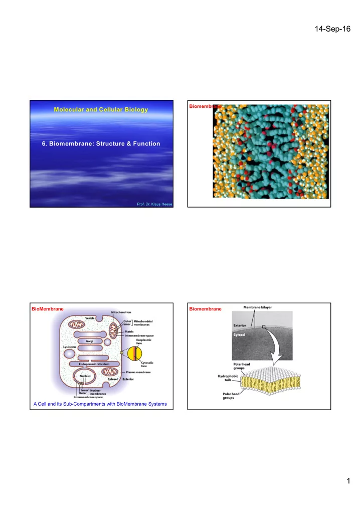

14-Sep-16 Biomembrane Molecular and Cellular Biology 6. Biomembrane: Structure & Function Prof. Dr. Klaus Heese BioMembrane Biomembrane A Cell and its Sub-Compartments with BioMembrane Systems 1
14-Sep-16 Biomembrane Biomembrane Molecules Molecules of a Biomembrane Molecules of a Biomembrane ---> fatty acid metabolism 2
14-Sep-16 Molecules of a Molecules of a Biomembrane Biomembrane PC = phosphatidylcholine; PE = phosphatidylethanol; PS = phosphatidylserine; PC = phosphatidylcholine; PE = phosphatidylethanol; PS = phosphatidylserine; SM = sphingomyelin; PI = phosphoinositol SM = sphingomyelin; PI = phosphoinositol Molecules of a Molecules of a Biomembrane Biomembrane Acetyl-CoA 3
14-Sep-16 Cholesterol § Plant and animal food contain sterols but only animal food contains cholesterol § Why? Cholesterol is made in the liver and plants do not have a liver § Cholesterol is needed to make bile, sex hormones, steroids and vitamin D. § It is the constituent of cell membrane structure § Dietary recommendation - <300 mg/d § Sources – egg yolks, liver, shellfish, organ foods Molecules of a Biomembrane Molecules of a Molecules of a Biomembrane Bio- Membrane 4
14-Sep-16 The Steroid Cholesterol has different effects on biomembrane fluidity at different temperatures Cholesterol Adding Cholesterol to a cell membrane reduces fluidity, therefore, making the cell membrane more rigid (c) Cholesterol within the animal cell membrane reducing phospholipid movement. Without cholesterol, cell membranes would be too fluid, not firm enough, and too permeable to some molecules. While cholesterol adds firmness and integrity to the plasma membrane and prevents it from becoming overly fluid, it also helps to maintain its fluidity. At the high concentrations as it is found in our cell's plasma membranes cholesterol helps to separate the phospholipids so that the fatty acid chains can't come together and crystallize. Therefore, cholesterol helps to prevent extremes-- whether too fluid, or too firm -- in the consistency of the cell membrane. PS Apoptosis Assay (lipid; PS) 5
14-Sep-16 Molecules of a Biomembrane – Membrane Proteins Cross-sectional views of the three structures formed by phospholipids in aqueous solutions The white spheres depict the hydrophilic heads of the phospholipids, and the squiggly black lines (in the yellow regions) represent the hydrophobic tails. Shown are a spherical micelle with a hydrophobic interior composed entirely of fatty acyl chains; a spherical liposome, which has two phospholipid layers and an aqueous center; and a two-molecule-thick sheet of phospholipids, or bilayer, the basic structural unit of bio-membranes. 6
14-Sep-16 Molecules of a Biomembrane – Membrane Proteins Molecules of a Biomembrane – Membrane Proteins 7
14-Sep-16 Biomembranes – Endo- / Exo-cytosis § Bulk transport across the plasma membrane occurs by exocytosis and endocytosis § Large proteins cross the membrane by different mechanisms Exocytosis • In exocytosis transport vesicles migrate to the plasma membrane, fuse with it, and release their contents (compare with synaptic (vesicle) neurotransmitter release) Endocytosis • In endocytosis the cell takes in macromolecules by forming new vesicles from the plasma membrane • (NGF uptake) Biomembranes - three types of endocytosis § Three types of endocytosis PHAGOCYTOSIS EXTRACELLULAR 1 µm CYTOPLASM FLUID In phagocytosis, a cell Pseudopodium engulfs a particle by Wrapping pseudopodia Pseudopodium around it and packaging of amoeba it within a membrane- enclosed sac large enough to be classified “ Food ” or as a vacuole. The other particle Bacterium particle is digested after the vacuole fuses with a Food lysosome containing vacuole Food vacuole hydrolytic enzymes. An amoeba engulfing a bacterium via phagocytosis (TEM). PINOCYTOSIS In pinocytosis, the cell 0.5 µm “ gulps ” droplets of extracellular fluid into tiny Plasma membrane vesicles. It is not the fluid Pinocytosis vesicles itself that is needed by the forming (arrows) in cell, but the molecules a cell lining a small dissolved in the droplet. blood vessel (TEM). Because any and all included solutes are taken into the cell, pinocytosis is nonspecific in the substances it transports. Vesicle 8
14-Sep-16 Retrograde NGF (Nerve Growth Factor) Signalling Receptor-mediated endocytosis enables the RECEPTOR-MEDIATED ENDOCYTOSIS cell to acquire bulk quantities of specific substances, even though those substances Coat protein may not be very concentrated in the Receptor Coated extracellular fluid. Embedded in the vesicle membrane are proteins with specific receptor sites exposed to the extracellular fluid. The receptor proteins are usually already clustered in regions of the membrane called coated pits, which are lined on their cytoplasmic Coated side by a fuzzy layer of coat proteins. pit Extracellular substances (ligands) bind Ligand to these receptors. When binding occurs, the coated pit forms a vesicle containing the ligand molecules. Notice that there are A coated pit relatively more bound molecules (purple) Coat inside the vesicle, other molecules and a coated protein (green) are also present. After this ingested vesicle formed during material is liberated from the vesicle, the receptor- receptors are recycled to the plasma mediated membrane by the same vesicle. endocytosis (TEMs). Plasma Cell signalling membrane 0.25 µm Molecular and Cellular Biology Relative permeability of a pure phospholipid bilayer to various molecules Transport of ions and small molecules across cell membranes A bilayer is permeable to small hydrophobic molecules and small uncharged polar Aquaporin, the water channel, consists of four identical transmembrane polypeptides molecules, slightly permeable to water and urea, and essentially impermeable to ions Prof. Dr. Klaus Heese and to large polar molecules. 9
14-Sep-16 Membrane Transport Proteins Gradients are indicated by triangles with the tip pointing toward lower concentration, electrical potential, or both. 1: pumps utilize the energy released by ATP hydrolysis to power movement of specific ions (red circles) or small molecules against their electrochemical gradient. 2: Channels permit movement of specific ions (or water) down their electrochemical gradient; they can also be controlled by e.g. ligand binding or phosphorylations etc : Transporters, which fall into three groups, facilitate movement of specific small molecules or ions. Uniporters transport a single type of molecule down its concentration gradient (3A). Cotransport proteins (symporters (3B) and antiporters (3C) catalyze the movement of one molecule against its concentration gradient (black circle), driven by movement of one or more ions down an electrochemical gradient (red circles). Differences in the mechanisms of transport by these three major classes of proteins account for their varying rates of solute movement. Transporters can also depend on ATP. Cellular uptake of glucose mediated by GLUT proteins exhibit simple Model of Uniport Transport by GLUT1 enzyme kinetics and greatly exceeds the calculated rate of glucose entry solely by passive diffusion In one conformation, the glucose-binding site faces outward; in the other, the binding site faces inward. Binding of The initial transport rate for the substrate S into the cell catalyzed by e.g. GLUT1: v=Vmax/(1+Km/[S]) glucose to the outward-facing site (step-1) triggers a conformational change in the transporter that results in the The initial rate of glucose uptake (measured as micromoles per milliliter of cells per hour) in the first few seconds is plotted against binding site facing inward toward the cytosol (step-2). Glucose then is released to the inside of the cell (step 3). increasing glucose concentration in the extracellular medium. In this experiment, the initial concentration of glucose in the cells is Finally, the transporter undergoes the reverse conformational change, regenerating the outward-facing binding always zero. Both, GLUT1, expressed by erythrocytes, and GLUT2, expressed by liver cells, greatly increase the rate of glucose site (step 4). If the concentration of glucose is higher inside the cell than outside, the cycle will work in reverse uptake (red and orange curves) at all external concentrations. Like enzyme-catalyzed reactions, GLUT-facilitated uptake of glucose (step-4 ---> step-1), resulting in net movement of glucose form inside to out. The actual confomrational changes exhibits a maximum rate (Vmax). The Km is the concentration at which the rate of glucose uptake is half maximal. GLUT2, with a Km are probably smaller than those depicted here. of about 20 mM, has a much lower affinity for glucose than GLUT1, with a Km of about 1.5 mM. 10
Recommend
More recommend