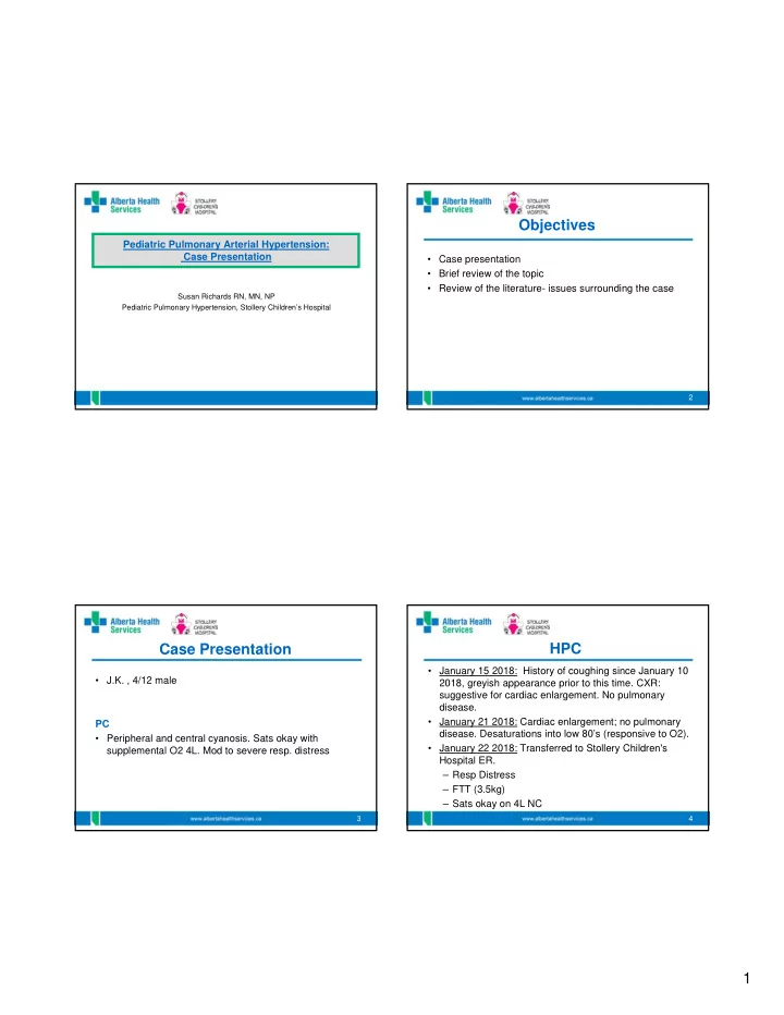

Objectives Pediatric Pulmonary Arterial Hypertension: Case Presentation • Case presentation • Brief review of the topic • Review of the literature- issues surrounding the case Susan Richards RN, MN, NP Pediatric Pulmonary Hypertension, Stollery Children’s Hospital 2 Case Presentation HPC • January 15 2018: History of coughing since January 10 • J.K. , 4/12 male 2018, greyish appearance prior to this time. CXR: suggestive for cardiac enlargement. No pulmonary disease. • January 21 2018: Cardiac enlargement; no pulmonary PC disease. Desaturations into low 80’s (responsive to O2). • Peripheral and central cyanosis. Sats okay with • January 22 2018: Transferred to Stollery Children's supplemental O2 4L. Mod to severe resp. distress Hospital ER. – Resp Distress – FTT (3.5kg) – Sats okay on 4L NC 3 4 1
PMH PMH • Medical HxP: • Pregnancy: Normal, no drugs taken – G6PC3 deficiency • Perinatal/Delivery: SVD, no complications, 2100grams (approx 4lbs 10 oz) – ASD • Postnatal: 24 hours post delivery cyanotic (Sats low – Undescended testicles 70%), RR 100, normal BS, septic workup, abx – Small for gestational age – 0.00 neutrophil – Osteopetrosis – WBC 0.8 – FTT – NP aspirate: + Human metapneumovirus • Med: G-CSF • Allergies: NKA • Immunizations: none 5 6 FH/SH O/E • General: Alert, irritable with examination, • First Child adequate head control. Wt 3.6kg /Ht: 59 cm • Caucasian • CNS: Anterior fontanelle soft • No one in family with known genetic defect • Resp: 40-50 mild in-drawing; suprasternal, tracheal tug. • No Hx of consanguinity Cry, squeaky but audible 2L O2 • Parents healthy (young) • CVS: WWP centrally; cool extremities.HR 109-157 SR;S2 split with increase P2; mild RV heave • Lives in a Hutterite Colony Rural Alberta • Abdomen: Liver 1 cm below RCM GU: undescended • No recent travel testes • Skin: superficial venous angiectasis 7 8 2
CXR ECG 9 10 ECHO Investigations • CXR: Cardiac Enlargement; lung and pleural spaces clear • ECG: SR, RAE, RVH, and LVH by voltage; towards RAD • Echocardiogram: small secundum ASD; bidirectional shunting RVSp 74 mmHg+Rap (SBP 98mmHg); PR mean PAp 32 mmHg; RVFAC 13%; small pericardial effusion. • Blood work: WBC 34.1; Neut.,30.2; ALT 72; NTproBNP 1624.0 pmol/L; Blood Culture,neg; NP aspirate;+ Human metapnemovirus &entero-rhinovirus; • MRSA, neg 11 12 3
Management Pro Beta Natriuretic Peptide NT proBNP • Echocardiogram assessing pulmonary veins. Test Number 1 2 3 4 5 • Start sildenafil 0.25 mg/kg x2 doses. Then 0.5 mg/kg x2 Collection Date 24-Jan-18 02-Feb-18 08-Feb-18 21-Feb-18 13-Mar-18 doses, and then 1 mg/kg per dose q8h pmol/L 1624.0 162.5 67.3 92.7 211.1 1800 • Aldactazide1mg/kg BID. 1600 1400 • Add in ETA 1200 1000 • Hematology to follow neutropenia 800 600 400 200 0 1 2 3 4 5 13 14 G6P Classification G6PC3 Pathophysiology General Points • Glucose-6-phosphatase enzyme is located in the endoplasmic reticulum (ER) and hydrolyses glucose-6- • Genetic disease autosomal recessive mutations on phosphate to glucose and phosphate. 17q21.31 • Loss of G6PC3 function shown to result in prevention of • Member of G6P family glucose recycling from the ER to the cytoplasm in the – G6PC1 (liver, gut and kidney) neutrophil – G6PC2 (pancreas) • Loss of G6PC3 activity also shown to increased • G6PC3 ubiquitously: Features congenital neutropenia, susceptibility to apoptosis in skin fibroblasts, superficial skin venous pattern heart and urogenital, neutrophils, and myeloid cells SHNL, FTT,PAH, cognitive impairment and/or endocrine abn 15 16 4
Pathophysiology Con’t G6PC3 Deficiency • Identification of the genetic basis of Dursun syndrome • Mutations in G6PC3 cause SCN4. adds to the existing knowledge that mutations in G6PC3 • Recurrent infections and prominent superficial venous can cause PPH. pattern are the most frequent clinical features • PPH is a known complication of type 1 glycogen • Congenital heart defects and urogenital malformations storage and establishing PPH as part of SCN4 • Dursun syndrome is triad of familial PPH, leucopenia, phenotype may suggests an important link between and ASD. glucose metabolism and PPH. • Dursun syndrome expands the pre-existing knowledge • Further studies needed of the phenotypic effects of mutations in G6PC3 • Should Dursun syndrome be considered as a subset of SCN4 with PPH as an important clinical feature? 17 18 Management/Challenges References • Treatment with G-CSF • Boztug, K.,et al. A syndrome with congenital neutropenia and mutations in G6PC3. New Eng. J. Med . 360: 32- 43, 2009. • Bacterial and viral infections • McDermott, D. H., et al. Severe congenital neutropenia resulting from G6PC3 deficiency with increased neutrophil CXCR4 expression and myelokathexis. Blood 116: 2793-2802, 2010. • Pulmonary vasodilator (s) • Arikoglu, T., et al. A novel G6PC3 gene mutation in severe congenital neutropenia: pancytopenia and variable bone marrow phenotype can also be part of this syndrome. European Journal of Hematology . 94: 79-82, 2014. • Banka, S. & Newman, W. A clinical and molecular review of ubiquitous glucose-6-phosphatase deficiency • Timing of images and invasive procedures caused by G6PC3 mutations. Journal of Rare Diseases . 8:84 1-17, 2013. • Banka, S., et al. Mutations in the G6PC3 Gene Cause Dursun Syndrome. American Journal of Medical Genetics . • Size of ASD 52A:2609–2611, 2010 • Lammers, A. et al. Diagnostics, monitoring and outpatient care in children with suspected pulmonary • No immunizations hypertension/paediatric pulmonary hypertensive vascular disease. Expert consensus statement on the diagnosis andtreatment of paediatric pulmonary hypertension. The European Paediatric Pulmonary Vascular Disease. • Medication compliance Heart BMJ . 102:ii1-ii13, 2016. • Kozlik-Feldmann, R., et al. Pulmonary hypertension in children with congenital heart disease (PAH-CHD, PPHVD-CHD). Expert consensus statement on the diagnosis and treatment of paediatric pulmonary • Multidisciplinary team approach hypertension. The European Paediatric Pulmonary Vascular Disease. Heart BMJ . 102:ii42-ii48, 2016. 19 20 5
Recommend
More recommend