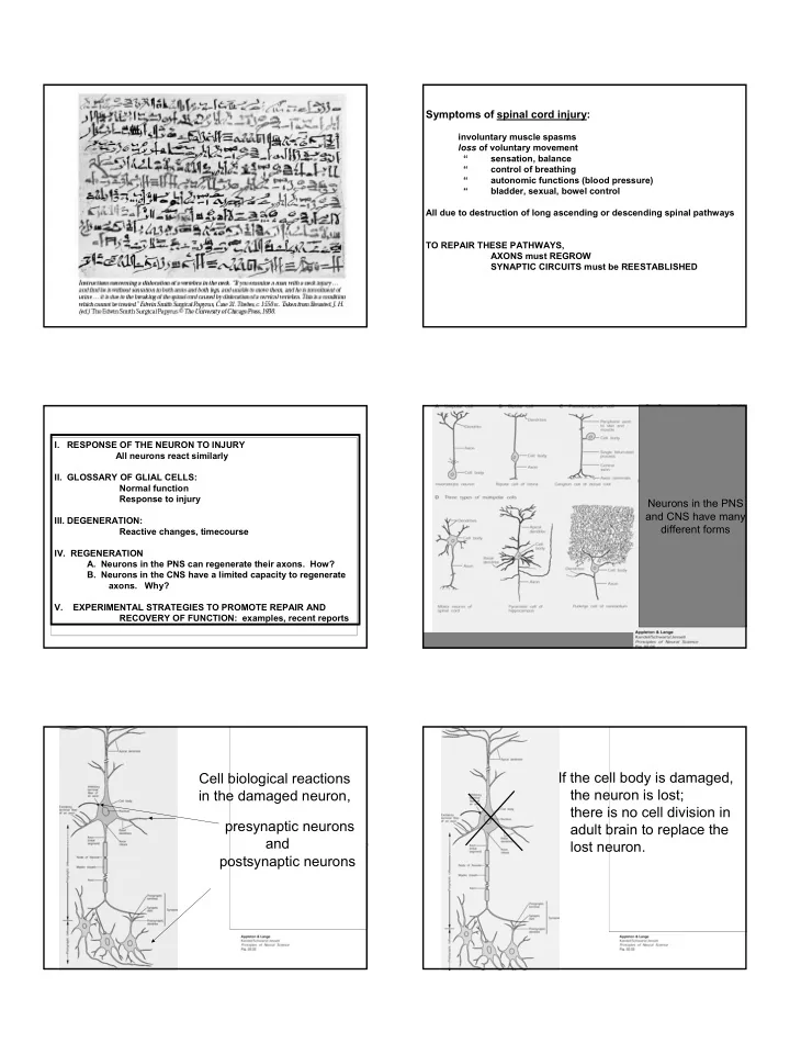

Symptoms of spinal cord injury: involuntary muscle spasms loss of voluntary movement “ sensation, balance “ control of breathing “ autonomic functions (blood pressure) “ bladder, sexual, bowel control All due to destruction of long ascending or descending spinal pathways TO REPAIR THESE PATHWAYS, AXONS must REGROW SYNAPTIC CIRCUITS must be REESTABLISHED I. RESPONSE OF THE NEURON TO INJURY All neurons react similarly II. GLOSSARY OF GLIAL CELLS: Normal function Response to injury Neurons in the PNS and CNS have many III. DEGENERATION: different forms Reactive changes, timecourse IV. REGENERATION A. Neurons in the PNS can regenerate their axons. How? B. Neurons in the CNS have a limited capacity to regenerate axons. Why? V. EXPERIMENTAL STRATEGIES TO PROMOTE REPAIR AND RECOVERY OF FUNCTION: examples, recent reports If the cell body is damaged, Cell biological reactions the neuron is lost; in the damaged neuron, there is no cell division in presynaptic neurons adult brain to replace the and lost neuron. postsynaptic neurons 1
I. RESPONSE OF THE NEURON TO INJURY (summary) If the axon is damaged, A. All neurons - despite different forms - react similarly the cell body is lost if the axon is severed close to the cell body, B. Principles but there is a chance that the axon will -If cell body damaged, the neuron dies, and is not replaced by cell division in mature brain. regenerate, even in the CNS. -If the axon is damaged or severed at a distance The postsynaptic, from the soma, there is a good chance of regeneration, primarily in the PNS. (and the presynaptic), neurons are also affected and may degenerate -CNS neurons have the capacity to regenerate. Types of glial cells I. RESPONSE OF THE NEURON TO INJURY 1. Myelin-forming: 2. Astrocytes a. Oligodendrocytes b. Schwann cells II. GLOSSARY OF GLIAL CELLS: Normal function, response to injury (CNS) (PNS) III. DEGENERATION: Signs, Timecourse IV. REGENERATION A. Neurons in the PNS can regenerate their axons. How? B. Neurons in the CNS have a limited capacity to regenerate axons. Why? V. EXPERIMENTAL STRATEGIES TO PROMOTE REPAIR AND RECOVERY OF FUNCTION: Principles, examples Myelin forming cells : (myelin important for conduction). oligodendroglia in CNS resting Schwann cells in PNS. oligodendrocytes (CNS) are inhibitory to axon regrowth in adult CNS regeneration; Schwann cells (PNS) are supportive, as a growth 3. Microglial cells surface and releaser of growth factors. Astroglia - activated development: supports axon growth and cell migration; mature: important for ion flux, synaptic function, blood-brain barrier injury: accumulate in scar, release excess matrix; inhibit axon growth? * phagocytic Microglia (resting) and macrophages (active) - cells of immune system, similar to monocytes. injury: help or hinder? ….not well-understood 2
REACTIONS TO INJURY WITHIN THE NEURON: I mmediately - I. RESPONSE OF THE NEURON TO INJURY 1. Synaptic transmission off 2. Cut ends pull apart and seal up, and swell, II. GLOSSARY OF GLIAL CELLS: Normal function, response to injury due to axonal transport in both directions III. DEGENERATION: Signs, Timecourse IV. REGENERATION A. Neurons in the PNS can regenerate their axons. How? B. Neurons in the CNS have a limited capacity to regenerate axons. Why? V. EXPERIMENTAL STRATEGIES TO PROMOTE REPAIR AND RECOVERY OF FUNCTION: Principles, examples REACTIONS TO INJURY WITHIN THE NEURON: I mmediately - 1. Synaptic transmission off 2. Cut ends pull apart and seal up, and swell, due to axonal transport in both directions Hours later - 3. Synaptic terminal degenerates - accumulation of NF, vesicles. 4. Astroglia surround terminal normally; after axotomy, astroglia interpose between terminal and target and cause terminal to be pulled away from postsynaptic cell. MINUTES after injury… -synaptic transmission off -cut ends swell Hours after injury….. Hours after injury….. SYNAPTIC TERMINAL DEGENERATES ASTROGLIA SURROUND SYNAPTIC TERMINAL Vesicles Synaptic accumulate neurofilaments NORMAL 3
REACTIONS TO INJURY WITHIN THE NEURON: I mmediately - 1. Synaptic transmission off 2. Cut ends pull apart and seal up, and swell, due to axonal transport in both directions Hours later - 3. Synaptic terminal degenerates - accumulation of NF, vesicles. 4. Astroglia suround terminal normally; after axotomy, interpose between terminal and target and cause terminal to be pulled away from postsynaptic cell. days - weeks - 5. Myelin breaks up and leaves debris (myelin hard to break down). 6. Axon undergoes Wallerian degeneration 7. Chromatolysis - cell body swells; nissl and nucleus eccentric. HOURS after… **If axon cut in PNS or CNS, changes are the same. synaptic terminal degenerates **The damaged neuron is affected by injury, as well as the pre- and postsynaptic neurons to it The damaged neuron is affected by injury Days to weeks after… as well as the neuron pre- and postsynaptic to it Binocular zone of right hemiretina Severing the axon causes degenerative changes in the injured neuron AND in the cells that have synaptic connections with the injured neuron. Fibers from the Monocular zone temporal retina* * project laterally Optic nerves Optic chiasm in the optic tract and Lateral geniculate Classically, degeneration of fibers and their targets terminate in layers 2,3,5 Optic nucleus tracts C I Dorsal has been used to trace neuronal circuits experimentally, of the Lateral Geniculate Nucleus C I and still is used to understand pathology post-mortem I C 6 5 Ventral 4 3 1 2 Magnocellular Parvocellular pathway pathway (M channel) (P channel) Primary visual cortex (area 17) Appleton & Lange Kandel/Schwartz/Jessell Principles of Neural Science Fig. 27.06 4
Binocular zone of right hemiretina I. RESPONSE OF THE NEURON TO INJURY II. GLOSSARY OF GLIAL CELLS: Normal function, response to injury Optic tract Monocular zone III. DEGENERATION: Signs, Timecourse, applications of “reading” trans-synaptic degeneration Optic nerves Optic chiasm IV. REGENERATION Laser lesion = lesion Lateral geniculate Optic nucleus A. Neurons in the PNS can regenerate their axons. How? tracts C (cat eye) = degeneration I Dorsal B. Neurons in the CNS have a limited capacity to regenerate C axons. Why? I Degenerating axons I C 6 (myelin stain) 5 Ventral 4 3 V. EXPERIMENTAL STRATEGIES TO PROMOTE REPAIR AND 1 2 The localization of degenerating fibers Magnocellular Parvocellular RECOVERY OF FUNCTION: Principles, examples pathway pathway (M channel) (P channel) can be used to trace where in the path Primary visual cortex the axons project, or where they terminate (area 17) Appleton & Lange Kandel/Schwartz/Jessell Principles of Neural Science Fig. 27.06 PNS neuron Regenerating axons form many sprouts, Reaction to injury some of which find Schwann cell tubes Axons sprout into Schwann cells -Ramon y Cajal Changes in the distal stump during degeneration and regeneration (PNS) 1 3 * Radioactive nerve growth factor Cut nerve stump 2 4 Macrophages clean debris, release mitogens for Schwann cells New Schwann cells form tubes, a conducive environment for growth: Schwann cells make laminin (growth-supportive extracellular matrix) Macrophages relase interleukin; interleukin stimulates Schwann cells to make Nerve Growth Factor * Nerve growth factor stimulates axon regeneration 5
Growth cone growth cones on regenerating Cell body axons: Growth in PNS I. RESPONSE OF THE NEURON TO INJURY II. GLOSSARY OF GLIAL CELLS: Normal function, response to injury IV. Neurons in the PNS can regenerate their axons. HOW? (summary) III. DEGENERATION: Signs, Timecourse a. After degeneration of distal axon and myelin, macrophages clean up debris. b. Macrophages release mitogens that induce Schwann cells to divide IV. REGENERATION A. Neurons in the PNS can regenerate their axons. How? c. The myelin-forming Schwann cells repopulate the nerve sheaths; B. Neurons in the CNS have a limited capacity to regenerate axons. d. Schwann cells make laminin Why? e. Macrophages make interleukin, which induces Schwann cells to make Nerve Growth Factor. V. EXPERIMENTAL STRATEGIES TO PROMOTE REPAIR AND RECOVERY OF FUNCTION: Principles, examples e. Axons sprout, and some sprouts enter new Schwann cell tubes f. Axonal growth cones successfully grow oligodendrocyte (in culture) B. Neurons in the mature CNS have a limited capacity to regenerate axons. WHY? PNS (or CNS) CNS axons can regrow, but… growth cone Growth is impeded by negative elements in the environment -extracellular matrix (laminin) is sparse; inhibitory proteoglycans increase -growth factors have different distributions compared to young brain Intracellular growth factors such as GAP-43 growth cone (important for intracellular signaling/growth cone advance) are low retracts 6
Recommend
More recommend