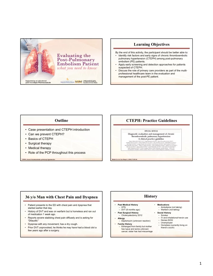

Learning Objectives By the end of this activity, the participant should be better able to: • Identify risk factors and early signs of chronic thromboembolic pulmonary hypertension (CTEPH) among post-pulmonary embolism (PE) patients. • Apply early screening and detection approaches for patients suspected of CTEPH. • Discuss the role of primary care providers as part of the multi- professional healthcare team in the evaluation and management of the post-PE patient. Outline CTEPH: Practice Guidelines • Case presentation and CTEPH introduction • Can we prevent CTEPH? • Basics of CTEPH • Surgical therapy • Medical therapy • Role of the PCP throughout this process CTEPH, chronic thromboembolic pulmonary hypertension Mehta S, et al. Can Respir J . 2010;17:301-34. History 36 y/o Man with Chest Pain and Dyspnea • Patient presents to the ED with chest pain and dyspnea that • Past Medical History • Medications – HTN – Amlodipine (not taking) started earlier that day. – DVT (6 months ago) – Warfarin (not taking) • History of DVT and was on warfarin but is homeless and ran out • Past Surgical History • Social History of medication 1 week ago. – Cholecystectomy 2012 – Smoker • Reports severe stabbing chest pain diffusely and is asking for – IV and inhalational heroin use • Allergy – Denies EtOH “Dilaudid.” – Daptomycin (unknown reaction) – Unemployed • Dyspnea with any movement; has a dry cough. • Family History – Homeless (currently living on – Estranged from family but mother • Prior DVT unprovoked, he thinks he may have had a blood clot a friend’s couch) has lupus and some unknown few years ago after a surgery. cancer, sister has had miscarriage 1
Physical Exam Temp BP HR RR 08/19 23:00 98.4/36.9 124/87 111 26 • General: NAD, alert & oriented × 4 • HEENT: OP clear, MMM, PERRL, EOMI • Neck: no LAD, no thyromegaly • CV: S1 and S2 normal, RRR, no m/r/g • Lungs: CTA b/l, no W/R/R, on 2L NC • Abdomen: s/nt/nd + bs, no HSM appreciated • Skin: track marks on upper extremities • Extremities: 2+ distal pulses, 1+ pitting edema to knee b/l • Neuro: CN II-XII intact NAD, no acute distress; HEENT, head, eyes, ears, nose, throat; OP, oropharynx; MMM, mucous membranes moist; PERRL, pupils equally round and reactive to light; EOMI, extra ocular muscles intact; LAD, lymphadenopathy; RRR, regular rate and rhythm; m/r/g, murmurs, rubs, gallops; CTA b/l, clear to auscultation bilaterally; W/R/R, wheezes/rales/rhonchi; s/nt/nd, soft/non-distended/non-tender; bs, bowel sounds; HSM, hepatosplenomegaly; CN, cranial nerve Acute and Chronic Complications of PE Risk Assessment in Acute PE • Acute PE incidence 100 per 100,000 patient years 1 – Increases with aging • Incidence has increased over time – Aging population and increased sensitivity of testing • Severity is widely variable 2 – Mortality rate in shock approaches 50% • Chronic Complications are common – PE recurrence in 40-50% at 10 years 3 – Significant morbidity and mortality with development of PH PE, pulmonary embolism; PH, pulmonary hypertension Konstantinides S.V., et al. J Am Coll Cardiol . 2016; 67(8):976-90. 1 Weiner RS, et al. Arch Intern Med . 2011; 171: 831-837. 2 Kucher N, et al. Circulation . 2006; 113: 577-582. Goldhaber, S.Z. Braunwald's Heart Disease, 84, 1681-1698. 3 Prandoni P, et al. Haematologica. 2007;92:199-205. Treatment of Acute PE RV Dysfunction in Acute PE • Obstruction of >30% of pulmonary vasculature correlates with • Low Risk → Anticoagulation RV dysfunction 1 – ACCP recommend NOAC rather than warfarin • 100% negative predictive value for PE-related death with • Less bleeding risk and greater convenience regards to RV dysfunction on TTE 2 – Duration of anticoagulation remains unclear • RV dysfunction associated with increased mortality 3 , though • Minimum 3 months, consider long term (24 months or more) low specificity on TTE • Unprovoked VTE has highest risk of recurrence • 24% ↑ risk of recurrent VTE with persistent RV dysfunction 4 RV, right ventricular; TTE, transthoracic echocardiogram; VTE, venous thromboembolism PE, pulmonary embolism; ACCP, American College of Chest Physicians; NOAC, novel oral anticoagulants; VTE, venous thromboembolism 1 Wolfe MW, et al. Am Heart J . 1994; 127: 1371-5. 2 Grifoni S, et al. Circulation . 2000; 101: 2817-22. Kearon C, et al. Chest . 2016;149:315-352. 3 Alpert JS, et al. JAMA . 1976; 236: 1477-80. 4 Grifoni S, et al. ACP J Club . 2007 Mar-Apr;146:46. 2
Treatment of Acute PE (cont.) • High Risk → Anticoagulation + Thrombolysis – 30-50% reduction in mortality with systemic thrombolysis, ~ 3 fold increase in major bleeding 1 – Catheter-directed thrombolysis and reperfusion • Appears efficacious, decreasing thrombus burden by > 50% 2 • Phase 3 trials are lacking and requires local expertise • Intermediate Risk → Anticoagulation + ? PE, pulmonary embolism 1 Chatterjee S, et al. JAMA . 2014;311:2414-2421. 2 Kuo WT, et al. Chest . 2015;148:667-673. Meyer G, et al. NEJM . 2014;370:1402-1411. Desai H, et al. Am J Med . 2017;130:e29-e32. What is Pulmonary Hypertension? • Diagnosed by RHC with mPAP ≥ 25 mm Hg Chronic Complications of PE: CTEPH – Normal mPAP ≤20 mm Hg at rest • Precapillary PH defined with PAWP ≤15 mm Hg – Normal ≤12 mm Hg • PAH defined by PVR >3 Woods units – (PVR = ∆Pressure/CO) – Normal PVR in some secondary PH RHC, right heart catheterization; mPAP, mean pulmonary arterial pressure; PH, pulmonary hypertension; PAWP, pulmonary arterial wedge pressure; PAH, pulmonary arterial hypertension; PVR, pulmonary vascular resistance; CO, cardiac output Hoeper MM, et al. J Am Coll Cardiol . 2013;62:D42-50. 3
Classification of Pulmonary Hypertension Group 4 Pulmonary Hypertension • Chronic thromboembolic PH • Estimated ~3% incidence after acute PE • Obstruction + arteriopathy • Treatment both surgical and medical Simonneau G, et al. J Am Coll Cardiol . 2013;62:D34-41. Lang IM, et al. Ann Am Thorac Soc . 2016;13(Suppl 3):S215-21. Can We Prevent CTEPH? Thrombolysis to Prevent CTEPH Fasullo S, et al. Am J Med Sci . 2011;341:33-9. Fasullo S, et al. Am J Med Sci . 2011;341:33-9. Thrombolysis to Prevent CTEPH Identification of CTEPH Fasullo S, et al. Am J Med Sci . 2011;341:33-9. 4
Incidence and Risk Factors of CTEPH in Detecting Patients After Acute PE CTEPH Yang S, et al. J Thorac Dis . 2015;7:1927-38. Galie N, et al. Eur Heart J . 2016;37:67-119. Monitoring for PH Following PE: Screen Appropriately The INFORM Study • Screening all patients 1 year after PE resulted in <1% diagnosis of CTEPH • Targeted screening has better yield Ende-Verhaar YM, et al. Thromb Res . 2017;151:1-7. Tapson VF, et al. Am J Med . 2016;129:978-985.e2. Monitoring for PH Following PE: V/Q Scan vs. CT Angiography The INFORM Study Patients with subsequent diagnosis of pulmonary hypertension • False positive V/Q in 15/149 – PAH, PVOD, PH with parenchymal lung disease • CT poor at identifying distal thromboembolic disease PVOD, pulmonary venoocclusive disease Tapson VF, et al. Am J Med . 2016;129:978-985.e2. Tunariu N, et al. J Nucl Med . 2007;48:680-4. 5
Patient returns 6 months later, reports dyspnea when V/Q Scan climbing 1 flight of stairs or ½ block on level ground. Our Patient Gets RHC HEMODYNAMIC DATA : Pressure (mm Hg) O 2 Saturations AORTA: 132/87 (105) 94% The Basics of CTEPH LV: 143 / 9 PCW: Poor quality tracing despite multiple attempts PA: 84/29 (49) 57.8% RV: 88/13 RA: 15 CARDIAC OUTPUT (L/MIN) by Thermodilution: 4.5; by Estimated Fick: 4.8 CARDIAC INDEX (L/MIN/M 2 ) by Thermodilution: 1.8; by Estimated Fick: 1.9 RESISTANCE (WOOD units = dynes-sec/cm 5 ) PULMONARY VASCULAR RESISTANCE (NL 20 ‒ 130) Thermodilution: 824 Estimated Fick: 771 LV, left ventricle; PCW, pulmonary capillary wedge; PA, pulmonary artery; RV, right ventricle; RA, right atrium CTEPH Pathogenesis of CTEPH Madani MM, et al. Ann Thorac Surg. 2012;94:97–103. Matthews DT, Hemnes AR. Pulm Circ . 2016;6:145–154. 6
Our Patient Gets Lab Results Risk Factors for CTEPH • Recurrent PE • Proximal disease • Antiphospholipid syndrome • Hemostatic Factors – Elevated Factor VIII, vW factor • Splenectomy – Erythrocytosis and thrombocytosis • Non-O blood group Kim NH, Lang IM. Eur Respir J . 2012;21:27-31. Pulmonary Endarterectomy • Performed through median sternotomy • Circulatory arrest for ~20 minutes at a time Therapies for CTEPH • Unilateral endarterectomy at each arrest • Can be successful to subsegmental branches • Jamieson Classification: – Type I – Acute or subacute proximally – Type II – Chronic disease proximally – Type III – Segmental and subsegmental only Pulmonary Endarterectomy Outcomes of PEA PEA, pulmonary endarterectomy Madani MM, et al. Ann Thorac Surg 2012;94:97–103. 7
Recommend
More recommend