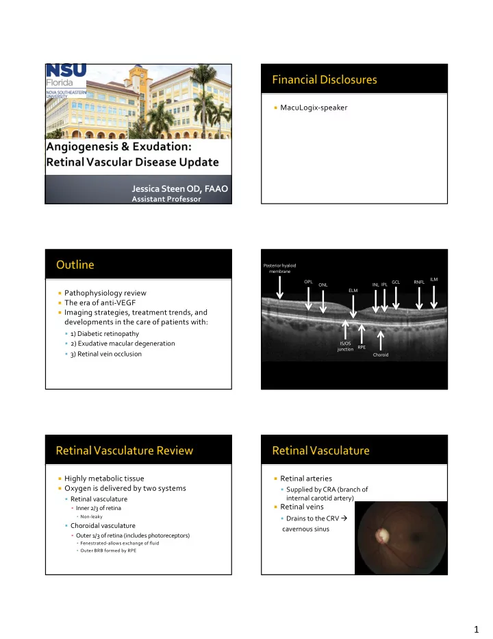

¡ MacuLogix-speaker Jessica Steen OD, FAAO Assistant Professor Posterior hyaloid membrane ILM OPL GCL RNFL ONL INL IPL ELM ¡ Pathophysiology review ¡ The era of anti-VEGF ¡ Imaging strategies, treatment trends, and developments in the care of patients with: § 1) Diabetic retinopathy § 2) Exudative macular degeneration IS/OS RPE junction § 3) Retinal vein occlusion Choroid ¡ Highly metabolic tissue ¡ Retinal arteries ¡ Oxygen is delivered by two systems § Supplied by CRA (branch of internal carotid artery) § Retinal vasculature ¡ Retinal veins ▪ Inner 2/3 of retina ▪ Non-leaky § Drains to the CRV à § Choroidal vasculature cavernous sinus ▪ Outer 1/3 of retina (includes photoreceptors) ▪ Fenestrated-allows exchange of fluid ▪ Outer BRB formed by RPE 1
¡ Retinal capillaries § 1) Superficial capillary plexus (GCL-to a lesser extent RNFL) ▪ Most affected in artery based conditions (HTN) § 2) Deep capillary plexus (INL) ▪ Prevenular capillary network ▪ Most affected in venous congestive disease (diabetes and RVO) ▪ Outer boundary is the outer plexiform layer § 3) Peripapillary capillary network ▪ Superficial, fewer anastomoses ¡ RPE § Loose attachment to PRs § Strong attachment to choriocapillaris and other RPE cells ¡ Choriocapillaris § Fed by posterior ciliary artery branches § Window defect (RPE defect) caused by choriocapillaris infarction Superficial: GCL Deep: INL ▪ Compartmentalized blood supply ¡ Choroid § Larger blood vessels, nerves, melanocytes, immune cells ▪ Presence of immunological cells represent source for inflammatory retinal disease 2
¡ The only thing that moves in the retina over ¡ En face flow formation and cross sectional time are red blood cells structural information ¡ Take the ‘difference’ between multiple B ¡ Not a replacement for FA/OCT scans at the same location to produce a § Provides new information ‘decorrelation signal’ ¡ Important in diagnosis of NV and macular ischemia ¡ Choroidal neovascularization ¡ Diagnosis of retinal vascular disease § Mactel type 2 § RVO ¡ Diabetic retinopathy § Microaneurysms, macular ischemia, NV ¡ AMD § Non-exudative lesions in eyes with intermediate AMD ¡ Neurodegenerative disease ¡ Open angle glaucoma Superficial retinal capillary plexus Deep retinal capillary plexus ¡ Static blood flow information § No leakage, pooling or staining ¡ Small field of view 3x3mm; 6x6mm; 8x8mm 3
¡ Motion artifacts are a big deal ¡ 1) Exudation § Loss of blood retinal barrier ¡ Sensitivity is a challenge in eyes with ▪ Accumulation of plasma fluid and lipid pathology ▪ Hard exudate and intraretinal edema ¡ 2) Ischemia § Capillary drop out leads to hypoxia ¡ Quantification of blood flow-not yet ▪ Microaneurysms, capillary drop out, collateral formation, neovascularization ¡ 3) Both ¡ End organ response to ¡ Type II: High incidence of DR at the time of systemic disease presentation ¡ Multifactorial condition § Annual exam ¡ Type I: No matter how poorly controlled, § Vascular component typically no retinopathy for 5-7 years § Hyperglycemic component ▪ Free-radical formation § Examine 5 years after diagnosis—or at age ten, then § Inflammation annually § Insulin-dependent type II patients are considered to § Compromised be of higher risk autoregulation ¡ Gestational DM ¡ Tissue damage to metabolically active sites § Do not seem to have increased risk of DR; no eye examination during pregnancy § Retina, kidney TABLE 1 D IABETIC R ETINOPATHY D ISEASE S EVERITY S CALE AND I NTERNATIONAL C LINICAL D IABETIC R ETINOPATHY D ISEASE S EVERITY S CALE ¡ Substratification Disease Severity Level Findings Observable upon Dilated Ophthalmoscopy No apparent retinopathy No abnormalities § Clusters 1-5 Mild NPDR (see Glossary) Microaneurysms only Moderate NPDR (see Glossary) More than just microaneurysms but less than severe NPDR ¡ Diabetic retinopathy most common in cluster Severe NPDR U.S. Definition Any of the following (4-2-1 rule) and no signs of proliferative retinopathy: 2 � Severe intraretinal hemorrhages and microaneurysms in each of four quadrants � Definite venous beading in two or more quadrants � Moderate IRMA in one or more quadrants § “Severe insulin-deficient diabetes” International Definition Any of the following and no signs of proliferative retinopathy: � More than 20 intraretinal hemorrhages in each of four quadrants ▪ Shares features of classic type I diabetes � Definite venous beading in two or more quadrants � Prominent IRMA in one or more quadrants ¡ Better, but-treat the patient, not the disease PDR One or both of the following: � Neovascularization � Vitreous/preretinal hemorrhage IRMA = intraretinal microvascular abnormalities; NPDR = nonproliferative diabetic retinopathy; PDR = proliferative diabetic retinopathy NOTE: � Any patient with two or more of the characteristics of severe NPDR is considered to have very severe NPDR. � PDR may be classified as high-risk and non-high-risk. See Table 6 for more information. Adapted with permission from Wilkinson CP, Ferris FL III, Klein RE, et al. Proposed international clinical diabetic retinopathy and diabetic macular edema disease severity scales. Ophthalmology 2003;110:1679. 4
¡ Vision loss occurs secondary to: ¡ Modified Airlie House-defined by § 1) Diabetic macular edema ETDRS in 1981 ¡ Very mild NPDR § MA only (level 20) § 2) Macular ischemia ¡ Mild NPDR § Hard exudate, cotton wool spots, § 3) Proliferative diabetic retinopathy and/or mild retinal hemorrhages (level 35) ¡ Leakage of lipoproteins from permeable ¡ Caused by microvascular occlusion or leakage capillaries ¡ ‘CSME’ defined by ETDRS § Hard exudate within 500μm of the center of the macula § Hard exudates at or within 500μm of the center of the macula with adjacent retinal thickening § Retinal thickening of 1DD of larger within 1DD of the center of the macula ¡ Color fundus photography ¡ Now, trend is an OCT-based classification § Great for documentation ¡ Fundus autofluorescence § Few indications that alter management ¡ FA § Evolved to be a test of retinal periphery ¡ ICG § Limited availability and utility ¡ OCT § THE most important ancillary test in retinal disease ¡ OCT angiography 5
Center-involved DME Non-center involved DME X Focal non-center involved DME TABLE 2 I NTERNATIONAL C LINICAL D IABETIC M ACULAR E DEMA D ISEASE S EVERITY S CALE Proposed Disease Severity Level Findings Observable upon Dilated Ophthalmoscopy Diabetic macular edema apparently absent No apparent retinal thickening or hard exudates in posterior pole Diabetic macular edema apparently present Some apparent retinal thickening or hard exudates in posterior pole If diabetic macular edema is present, it can be categorized as follows: Proposed Disease Severity Level Findings Observable upon Dilated Ophthalmoscopy * Diabetic macular edema present � Mild diabetic macular edema: some retinal thickening or hard exudates in posterior pole but distant from the center of the macula � Moderate diabetic macular edema: retinal thickening or hard exudates approaching the center of the macula but not involving the center � Severe diabetic macular edema: retinal thickening or hard exudates involving the center of the macula Reproduced with permission from Wilkinson CP, Ferris FL III, Klein RE, et al. Proposed international clinical diabetic retinopathy and diabetic macular edema disease severity scales. Ophthalmology 2003;110:1680. * Hard exudates are a sign of current or previous macular edema. Diabetic macular edema is defined as retinal thickening; this requires a three-dimensional assessment that is best performed by dilated examination using slit-lamp biomicroscopy and/or stereoscopic fundus photography. Optical coherence tomography may supplement the fundus evaluation for determining the presence of diabetic macular edema. ¡ Early clinical feature of non-proliferative ¡ Weakening of diabetic retinopathy capillary wall ¡ Large MAs visible § Thickening of basement membrane, pericyte loss, clinically MAs, increased permeability ▪ Leads to loss of vessel perfusion, hypoxia, increased ¡ Leak VEGF, neovascularization § Cause intraretinal § Most commonly found in the inner nuclear layer edema ▪ Generally surround areas of capillary loss ▪ Deep capillary plexus 6
¡ Deep retinal hemorrhages § Inner nuclear layer, outer plexiform layer, outer nuclear layer § From pre-venular capillaries ▪ DM, RVO § Represent ruptured microaneurysms ▪ Do not leak on FA ¡ Cotton wool spot § Really not an “infarct” 7
Recommend
More recommend