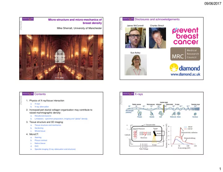

09/06/2017 Micro-structure and micro-mechanics of Disclosures and acknowledgements breast density James McConnell Charles Streuli Mike Sherratt, University of Manchester Sue Astley Contents X-rays 1. Physics of X-ray/tissue interaction a. X-rays b. X-ray attenuation 2. Increased peri-ductal collagen organisation may contribute to raised mammographic density: a. Results/conclusions b. Limitations - specimen preparation, imaging and “global” density 3. Tissue structure and 3D imaging: a. Tissue structure and mechanics. b. Sectioning c. Whole tissue 4. MicroCT: a. Staining b. Phase contrast c. Native tissue d. DVC e. Speckle imaging (X-ray attenuation and structure) 1
09/06/2017 X-ray attenuation Why should breast tissue induce X-ray contrast? Attenuation: Scattering: Elastic Inelastic Absorption: Photoelectric Probability of photoelectric absorption: Beer Lambert law: Patient cohort Contents 1. Physics of X-ray/tissue interaction a. X-rays Study cohort b. X-ray attenuation 2. Increased peri-ductal collagen organisation may contribute to raised mammographic density: a. Results/conclusions b. Limitations - specimen preparation, imaging and “global” density 3. Tissue structure and 3D imaging: a. Tissue structure and mechanics. b. Sectioning c. Whole tissue 4. MicroCT: a. Staining b. Phase contrast c. Native tissue d. DVC e. Speckle imaging (X-ray attenuation and structure) 2
09/06/2017 Quantifying collagen – by picrosirious red and Quantifying collagen – by Masson’s trichrome polarised light Masson’s trichrome Quantifying collagen – by picrosirious red and High MD tissue is characterised by large collagen polarised light bundles. Collagen organisation - AFM 3
09/06/2017 Collagen can also be too organised Mass spectrometry suggest that collagen organising proteins (i.e. periostin and collagen XVI) mediate local stiffness Contents 1. Physics of X-ray/tissue interaction a. X-rays b. X-ray attenuation 2. Increased peri-ductal collagen organisation may contribute to Local tissue organisation, mechanics and proteomics raised mammographic density: a. Results/conclusions correlates with b. Limitations - specimen preparation, imaging and “global” density 3. Tissue structure and 3D imaging: Global tissue X-ray density a. Tissue structure and mechanics. Local tissue X-ray density b. Sectioning c. Whole tissue 4. MicroCT: a. Staining b. Phase contrast c. Native tissue d. DVC e. Speckle imaging (X-ray attenuation and structure) 4
09/06/2017 The mechanical behaviour of ECM-rich tissues is Aberrant ECM structure and disease determined by composition and structure Cells Elastic fibres Fibrillar collagens Articular Structural Skin cartilage Proteoglycans Network collagens Adhesive glycoproteins Soluble Elastic Cardiac arteries muscle Proteases/ Cytokines inhibitors Danielson et al., 1997 J. Cell Biol. 136:729-743. Decorin KO mice lack collagen organisation Decorin “decorates” collagen fibrils Danielson et al., 1997 J. Cell Biol. 136:729-743. 5
09/06/2017 Tissues are inherently three dimensional 2D sections: sampling issues and sectioning artefacts Hole Disrupted adventitia Intact 3D media Tissues Hole Intact adventitia 50µm 2D sections 2D sections: sampling issues and sectioning artefacts Disrupted stratum corneum Serial Block face Confocal X-ray Holes sectioning imaging microscopy tomography Epidermis Dermis 50µm Holes 6
09/06/2017 Serial sectioning and reconstruction…..ancient Serial sectioning and reconstruction……and modern Serial block face imaging Confocal microscopy 7
09/06/2017 The ideal imaging technique would enable….. X-ray imaging 1. Physics of X-ray/tissue interaction a. X-rays b. X-ray attenuation 2. Increased peri-ductal collagen organisation may contribute to …..rapid, high contrast, 3D visualisation and raised mammographic density: mechanical characterisation of large volumes of a. Results/conclusions opaque native tissues at sub-micrometer length scales b. Limitations - specimen preparation, imaging and “global” density 3. Tissue structure and 3D imaging: a. Tissue structure and mechanics. b. Sectioning c. Whole tissue 4. MicroCT: a. Staining b. Phase contrast c. Native tissue d. DVC e. Speckle imaging (X-ray attenuation and structure) Radiography Tomography Principals of Micro-Computed X-Ray Tomography X-ray imaging facilities (microCT or sometimes µCT) 8
09/06/2017 Principals of microCT Specimen preparation artefacts Phase contrast MicroCT resolves major recognisable structures in intact arteries Paraffin Medial layer Adventitial layer 9
09/06/2017 Segmentation of medial and adventitial layers across Arterial pressurisation apparatus 100’s of slices Artery mounted in bath between two glass cannulas A pressure servo and peristaltic pump maintain the artery at a set isobaric Myograph arms rotate the pressure assembly into a bath of fixative or Liquid N2-cooled pentane. Medial but not adventitial volume is maintained in Pressure profoundly affects 3D vessel structure pressurised vessels Intimal & adventitial surface topography 0mmHg 25 110mmHg Measurements from 1 rat, 450 0mmHg slices (338 µm) 110mmHg 65258µm 2 20 (SD 1114µm 2 ) Frequency (%) Mann Whitney U test 15 66962µm 2 p<0.0001 (SD 1603µm 2 ) 10 5 Lumen volume 0 50000 60000 70000 80000 Media CSA (microns^2) Elastic lamellae 20 0mmHg tortuosity / 18 56844µm 2 110mmHg (SD 1750µm 2 ) 16 spacing / 14 Frequency (%) thickness 12 72658µm 2 10 (SD 4855µm 2 ) 8 6 4 2 0 Medial and adventitial 50000 60000 70000 80000 Adventitia CSA (microns^2) layer thickness 10
09/06/2017 Breast microCT – preliminary scans Breast microCT – preliminary scans Specimen fixation artefacts 4µm 11
09/06/2017 The ideal imaging technique would enable….. Synchrotron? …..rapid, high contrast, 3D visualisation and mechanical characterisation of large volumes of opaque native tissues at sub-micrometer length scales But scans on laboratory instruments take 10 hours. Therefore we need: A higher X-ray flux Which means a bigger machine Which means a synchrotron Synchroton imaging of native tissue Synchroton imaging of native tissue 12
09/06/2017 Synchrotron imaging and Synchrotron imaging and DVC digital volume correlation (DVC) Synchrotron microCT of sequentially compressed DVC and 3D strain maps disc Slice 880 13
09/06/2017 Speckle imaging: simultaneous absorption, DVC and 3D strain maps scatteing and phase contrast 1. MicroCT: a. Staining b. Phase contrast c. Native tissue d. DVC e. Speckle imaging (X-ray attenuation and structure) Absorption Dark field (scattering) Horizontal phase Vertical phase Combined phase Summary Public awareness 1. Physics of X-ray/tissue interaction X-ray contrast due to scattering and absorption 2. Increased peri-ductal collagen organisation may contribute to raised mammographic density: Raised MD correlated with mechanically stiff collagen fibril bundles 3. Tissue structure and 3D imaging: Tissue and molecular structure is a key mediator of mechanical properties 3D structure can be characterised by sectioning or confocal methods 4. MicroCT: Image large volumes of native tissue at micron-resolutions Resolve micro-structure and micro-calcifictions in breast tissue Map 3D strain May be able to simultaneously characterise tissue structure and X-ray attenuation 14
09/06/2017 Public awareness Acknowledgements 15
Recommend
More recommend